KNOWLEDGE BAG OF BIOCHEMISTRY

Tuesday 29 September 2015

NUCLEAR FUSION ON SMALL SCALE MAY BE A NEW ENERGY CREATOR......

Fusion energy may soon be used in small-scale power stations. This means producing environmentally friendly heating and electricity at a low cost from fuel found in water. Both heating generators and generators for electricity could be developed within a few years, according to research that has primarily been                conducted at the University of Gothenburg.


Nuclear fusion is a process whereby atomic nuclei melt together and release energy. Because of the low binding energy of the tiny atomic nuclei, energy can be released by combining two small nuclei with a heavier one. A collaboration between researchers at the University of Gothenburg and the University of Iceland has been to study a new type of nuclear fusion process. This produces almost no neutrons but instead fast, heavy electrons (muons), since it is based on nuclear reactions in ultra-dense heavy hydrogen (deuterium).
"This is a considerable advantage compared to other nuclear fusion processes which are under development at other research facilities, since the neutrons produced by such processes can cause dangerous flash burns," says Leif Holmlid, Professor Emeritus at the University of Gothenburg.
No radiation The new fusion process can take place in relatively small laser-fired fusion reactors fueled by heavy hydrogen (deuterium). It has already been shown to produce more energy than that needed to start it. Heavy hydrogen is found in large quantities in ordinary water and is easy to extract. The dangerous handling of radioactive heavy hydrogen (tritium) which would most likely be needed for operating large-scale fusion reactors with a magnetic enclosure in the future is therefore unnecessary.
" A considerable advantage of the fast heavy electrons produced by the new process is that these are charged and can therefore produce electrical energy instantly. The energy in the neutrons which accumulate in large quantities in other types of nuclear fusion is difficult to handle because the neutrons are not charged. These neutrons are high-energy and very damaging to living organisms, whereas the fast, heavy electrons are considerably less dangerous."
Neutrons are difficult to slow down or stop and require reactor enclosures that are several meters thick. Muons -- fast, heavy electrons -- decay very quickly into ordinary electrons and similar particles.
Research shows that far smaller and simpler fusion reactors can be built. The next step is to create a generator that produces instant electrical energy.
The research done in this area has been supported by GU Ventures AB, the holding company linked to the University of Gothenburg. The results have recently been published in three international scientific journals.

Monday 28 September 2015

SYNTHETIC BLOOD: Deep-diving Whales Answered it........

The ultra-stable properties of the proteins that allow deep-diving whales to remain active while holding their breath for up to two hours biochemist to finish a 20-year quest to create lifesaving synthetic blood for human trauma patients.


The researchers compared the muscle protein myoglobin from humans, whales and other deep-diving mammals. Myoglobin holds oxygen for ready use inside muscle cells, and the study found that marine mammals have ultra-stable versions of myoglobin that tend not to unfold. The researchers found that stability was the key for cells to make large amounts of myoglobin, which is explains why deep-diving mammals can load their muscle cells with far more myoglobin than humans.
"Whales and other deep-diving marine mammals can pack 10-20 times more myoglobin into their cells than humans can, and that allows them to 'download' oxygen directly into their skeletal muscles and stay active even when they are holding their breath," said Olson, Rice's Ralph and Dorothy Looney Professor of Biochemistry and Cell Biology. "The reason whale meat is so dark is that it's filled with myoglobin that is capable of holding oxygen. But when the myoglobin is newly made, it does not yet contain heme. We found that the stability of heme-free myoglobin is the key factor that allows cells to produce high amounts of myoglobin."
That's important to Olson because he wants to create a strain of bacteria that can generate massive quantities of another protein that's closely related to myoglobin. Olson has spent two decades studying hemoglobin, a larger, more complex oxygen-carrying protein in blood. Olson's goal is to create synthetic blood for use in transfusions. Hospitals and trauma specialists currently rely on donated whole blood, which is often in short supply and has a limited storage life. A crucial part of Olson's plan is maximizing the amount of hemoglobin that a bacterium can express.
"Our results confirm that protein stability is the key," Olson said. "In this study, Premila and George developed an in vitro method for testing myoglobin expression outside of living cells. That allowed us to carefully control all the variables. We found that the amount of fully active myoglobin expressed was directly and strongly dependent on the stability of the protein before it bound the heme group."
All proteins have a characteristic shape, and the globin family of proteins is shaped around a pocket where heme is stored. The heme pocket opens and closes -- much like the pocket of a baseball glove -- to trap and release oxygen.
Samuel, a graduate student in the Department of BioSciences at Rice, said the heme-free form of myoglobin that she studied is called apoprotein or apomyoglobin.
"The more stable the apoprotein, the more final product we could make," she said. "Human apomyoglobin isn't very stable at all compared to that of the diving mammals, which have versions of the apoglobin that are up to 60 times more stable than ours."
Samuel said the stability differences aren't obvious if one simply compares the overall structures of the myoglobin from each species. Their overall shapes, including the shapes of their heme pockets, are the same. However, thanks to subtle differences in their amino acid sequences, the more stable myoglobins are better able to retain their shapes. Samuel said this underlying stability only becomes apparent when one studies the heme-free, or "apo" versions of the protein. She measured stability using chemicals that forced the apoproteins to unfold. By carefully measuring the amount of chemical required, she was able to precisely measure stability.
She said her work was made possible by three earlier studies. In 1999, Emily Scott, a graduate student in Olson's lab, noticed that sperm whale apomyoglobin was much more resistant to chemically induced unfolding than the corresponding human or pig apoproteins. Scott wondered if the resistance to unfolding was a trait of deep-diving whales, so she gathered samples from a variety of mammals and confirmed the idea in 2000.
At the same time, study co-author Smith, another of Olson's graduate students, was examining a catalog of 250 mutant sperm-whale apomyoglobins. He noticed that a certain class of mutations in the heme pocket caused the proteins to become extraordinarily stable even though the mutations damaged their ability to bind heme and oxygen.
Finally, in 2013, Michael Berenbrink of Liverpool University and Kevin Campbell of the University of Manitoba noted that deep-diving mammals expressed large amounts of myoglobin in their muscle tissue. Berenbrink and Campbell systematically analyzed the genes and available information for all mammalian myoglobins, including those from deep-diving species, and found that the myoglobins from aquatic mammals had large positive surface charges compared with those from land animals. They hypothesized that the charge differences allowed the aquatic species to pack more myoglobin into their muscle cells.
"I heard Berenbrink present his work, and I wondered whether we should re-investigate Emily's and Lucian's work on expression levels and apoglobin stability," Olson said. "At the time, we were in the process of trying to screen large-scale libraries of hemoglobin mutants to try to select for higher stability and expression as part of our work on evaluating blood substitutes. George had suggested we use a wheat-germ-based cell-free translation system for those screens, and Premila was preparing to test the methods with myoglobin.
"The three of us decided she should conduct her tests on a series of proteins that included myoglobins from humans, pigs and several of the deep-diving mammals that Emily had tested and Berenbrink had examined," Olson said. "We also used Lucian's Ph.D. results to construct three mutants that were far more stable than anything found in nature."
In her tests, Samuel compared the stability and cell-free expression level of myoglobins from humans, pigs, goosebeak whales, gray seals, sperm whales, dwarf sperm whales and the three mutants, which had low heme affinity but were 50 times more stable than apomyoglobins from the whales. The research confirmed that the stability of apoprotein is directly correlated with expression levels. For example, very little pig and human myoglobin could be made in the cell-free system, which yielded 10- to 20-fold higher amounts of whale and mutant myoglobins.
The results of the cell-free study unequivocally verify the expression-stability correlations that had been anecdotally observed in previous work in both mammalian cells and E. coli, Olson said.
"This work is very important for our projects on synthetic blood substitutes and determining the toxicity of acellular hemoglobin," he said. "Premila has laid the groundwork for high-throughput screening of large libraries of hemoglobin variants without the need for purifying milligram quantities of pure protein. This method is a big step forward in our efforts to identify more stable recombinant hemoglobins."

Saturday 26 September 2015

WHAT PROTEINS POWERS THE PUMPING HEART???

Heart Researchers have uncovered a treasure trove of proteins, which hold answers about how our heart pumps -- a phenomenon known as contractility. These molecules haven't been studied in the heart and little is known about what they do in other tissues, the investigators says......


The Department of Medical Biophysics, the team used high-throughput methods to identify more than 500 membrane proteins on the surfaces of cardiac contractile cells, which are likely to have a critical role in normal heart function. The proteins may also play a part in heart failure and abnormal heartbeat patterns known as arrhythmias.
"In addition to providing a new understanding of what makes our hearts pump, these findings could also help researchers uncover new information about how heart disease affects the signal pathways in our hearts. That might pave the way to find ways to prevent or reverse those changes," says Gramolini.
During the study, the researchers found about 500 novel molecules that have been conserved throughout evolution. These molecules haven't been studied in the heart and little is known about what they do in other tissues.
The group's research focused on a protein called transmembrane protein 65 (Tmem65). By studying human stem cells and zebrafish using cell imaging and biochemical techniques, the researchers discovered that Tmem65 is involved in communication and electrical processes known as electrical coupling and calcium signaling. The team showed that Tmem65 regulates the connection point between adjacent cardiac contractile cells where it contributes to making the heart contract normally. Removing the protein had fatal consequences. The team also identified Tmem65 as the first critical tool for stem-cell researchers to monitor the maturation of cells in the heart's two main chambers, known as ventricles.

"These proteins are theoretically targetable for intervention as well as basic study. In this study, our focus was on Tmem65, but there are 555 proteins that we identified and showed that they are present throughout many species and are conserved throughout evolution-- at least in the mouse and the human -- in the heart's membrane-enriched contractile cells. Tmem65 was only the number-one candidate in our study, but theoretically, we have 554 other proteins to work through," says Gramolini.
The study, published in Nature Communications, also provides the first resource of healthy human and mouse heart-cell proteins that will help scientists develop a better understanding the mechanisms involved in cardiac disease.
Gramolini says the findings are essential for understanding cardiac biology and hopes they open the door for further study into health and disease in his lab and others.
"We need to figure out what all of these molecules are doing. My team and I hope our research sets the stage for other people to begin to pick up some of this work," says Gramolini. "These are molecules that haven't been studied, but must play some role in heart function. If a protein is conserved in evolution, generally it must have a critical function. We are very excited to look at the role of a number of these new proteins."

Friday 25 September 2015

NOW VIRUSES FIGHT AGAINST HARMFUL BACTERIA

Engineered viruses could combat human disease and improve food safety.


In the hunt for new ways to kill harmful bacteria, scientists have turned to a natural predator: viruses that infect bacteria. By tweaking the genomes of these viruses, known as bacteriophages, researchers hope to customize them to target any type of pathogenic bacteria.


To help achieve that goal, MIT biological engineers have devised a new mix-and-match system to genetically engineer viruses that target specific bacteria. This approach could generate new weapons against bacteria for which there are no effective antibiotics, says Timothy Lu, an associate professor of electrical engineering and computer science and biological engineering.
"These bacteriophages are designed in a way that's relatively modular. You can take genes and swap them in and out and get a functional phage that has new properties," says Lu, the senior author of a paper describing this work in the Sept. 23 edition of the journal Cell Systems.
These bacteriophages could also be used to "edit" microbial communities, such as the population of bacteria living in the human gut. There are trillions of bacterial cells in the human digestive tract, and while many of these are beneficial, some can cause disease. For example, some reports have linked Crohn's disease to the presence of certain strains of E. coli.
"We'd like to be able to remove specific members of the bacterial population and see what their function is in the microbiome," Lu says. "In the longer term you could design a specific phage that kills that bug but doesn't kill the other ones, but more information about the microbiome is needed to effectively design such therapies."
The paper's lead author is Hiroki Ando, an MIT research scientist. Other authors are MIT research scientist Sebastien Lemire and Diana Pires, a research fellow at the University of Minho in Portugal.


Customizable viruses
The Food and Drug Administration has approved a handful of bacteriophages for treating food products, but efforts to harness them for medical use have been hampered because isolating useful phages from soil or sewage can be a tedious, time-consuming process. Also, each family of bacteriophages can have a different genome organization and life cycle, making it difficult to engineer them and posing challenges for regulatory approval and clinical use.
The MIT team set out to create a standardized genetic scaffold for their phages, which they could then customize by replacing the one to three genes that control the phages' bacterial targets.
Many bacteriophages consist of a head region attached to a tail that enables them to latch onto their targets. The MIT team began with a phage from the T7 family that naturally killsEscherichia coli. By swapping in different genes for the tail fiber, they generated phages that target several types of bacteria.
"You keep the majority of the phage the same and all you're changing is the tail region, which dictates what its target is," Lu says.
To find genes to swap in, the researchers combed through databases of phage genomes looking for sequences that appear to code for the key tail fiber section, known as gp17.
After the researchers identified the genes they wanted to insert into their phage scaffold, they had to create a new system for performing the genetic engineering. Existing techniques for editing viral genomes are fairly laborious, so the researchers came up with an efficient approach in which they insert the phage genome into a yeast cell, where it exists as an "artificial chromosome" separate from the yeast cell's own genome. During this process the researchers can easily swap genes in and out of the phage genome.
"Once we had that method, it allowed us very easily to identify the genes that code for the tails and engineer them or swap them in and out from other phages," Lu says. "You can use the same engineering strategy over and over, so that simplifies that workflow in the lab."
A targeted strike
In this study, the researchers engineered phages that can target pathogenic Yersinia and Klebsiella bacteria, as well as several strains of E. coli. These are all part of a group known as Gram-negative bacteria, against which there are few new antibiotics. This group also includes microbes that can cause respiratory, urinary, and gastrointestinal infections, including pneumonia, sepsis, gastritis, and Legionnaires' disease.
One advantage of the engineered phages is that unlike many antibiotics, they are very specific in their targets. "Antibiotics can kill off a lot of the good flora in your gut," Lu says. "We aim to create effective and narrow-spectrum methods for targeting pathogens."

Tuesday 22 September 2015

PROTEIN ASSEMBLE, DISASSEMBLE ON COMMAND....

Gene sequences may enable control of building bio-structures.


Scientists have deciphered the genetic code that instructs proteins to either self-assemble or disassemble in response to environmental stimuli, such as changes in temperature, salinity or acidity. The discovery provides a new platform for drug delivery systems and an entirely different view of cellular functions.


The advance was made by researchers at Duke University and is the first time that scientists have reported the ability to create biological structures that are readily programmed to assemble and disassemble. With this knowledge in hand, researchers have opened a new world for designer proteins and investigations into nanotechnology, biotechnology and medical treatments.
The study appears September 21 in Nature Materials.
"The very simple design rules that we have discovered provide a powerful engineering tool for many biomedical and biotechnology applications," said Ashutosh Chilkoti, chair of the Department of Biomedical Engineering at Duke. "We can now, with a flick of a switch and a temperature jump, make a huge range of biological molecules that either assemble or disassemble."
The study investigated several triggers that can cause protein structures to assemble or break apart, but it primarily focused on heat. Protein-based structures that self-assemble when heated and remain stable inside of the bloodstream have long been used in a variety of applications. The opposite behavior, however, has long eluded researchers, especially outside of the carefully controlled environment of a chemistry lab.
"Nobody has been able to make these kinds of materials with the degree of complexity that we have now demonstrated," said Felipe Garcia Quiroz, a former graduate student in Chilkoti's laboratory and first author of the new study.
Chilkoti's lab has designed self-assembling proteins for drug delivery systems for several years. Simply by adding heat, these new packing materials put themselves together and help control where and when drugs are released inside the body through non-temperature-related mechanisms such as changes in acidity levels.
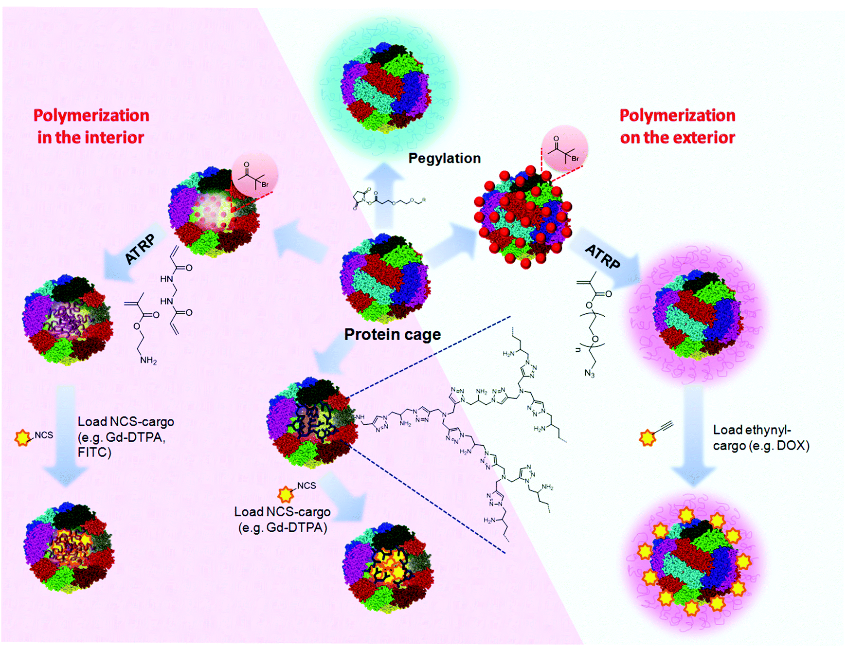
With the new discovery, however, drugs could be encapsulated in protein cages that accumulate inside of a tumor and dissolve once heated. Not only would this provide a more accurate way of delivering drugs, but the cages themselves could be used therapeutically.
"These packaging systems have always been inert, but now we can make these materials from bioactive components," said Quiroz. "Once the cages get there and deliver their cargo, they could break down into additional therapeutic agents. We can now design two things into one."
The research also provides new insights into the everyday functions of cells. Because the laboratory identified the genetic sequences that encode this behavior, they were able to point out a long list of human proteins that likely exhibit it.
"This paper shows the incredible richness of peptide sequences that already have this very simple switch," said Chilkoti.
Why they assemble and what function it serves, however, remains an open question.
"These findings will be exciting to both the materials science and the biochemistry communities," said Quiroz. "They'll be able to push the limits of what we know about these kinds of materials and then go back to explore how biology is already making use of them."

Targeting DNA: PROTEIN-SENSOR COULD DETECT VIRAL INFECTION AND KILL CANCER CELLS...

MIT biological engineers have developed a modular system of proteins that can detect a particular DNA sequence in a cell and then trigger a specific response, such as cell death.

This system can be customized to detect any DNA sequence in a mammalian cell and then trigger a desired response, including killing cancer cells or cells infected with a virus, the researchers say.
"There is a range of applications for which this could be important," says James Collins, the Termeer Professor of Medical Engineering and Science in MIT's Department of Biological Engineering and Institute of Medical Engineering and Science (IMES). "This allows you to readily design constructs that enable a programmed cell to both detect DNA and act on that detection, with a report system and/or a respond system."
Collins is the senior author of a Sept. 21 Nature Methods paper describing the technology, which is based on a type of DNA-binding proteins known as zinc fingers. These proteins can be designed to recognize any DNA sequence.
"The technologies are out there to engineer proteins to bind to virtually any DNA sequence that you want," says Shimyn Slomovic, an IMES postdoc and the paper's lead author. "This is used in many ways, but not so much for detection. We felt that there was a lot of potential in harnessing this designable DNA-binding technology for detection."


Sense and respond
To create their new system, the researchers needed to link zinc fingers' DNA-binding capability with a consequence -- either turning on a fluorescent protein to reveal that the target DNA is present or generating another type of action inside the cell.
The researchers achieved this by exploiting a type of protein known as an "intein" -- a short protein that can be inserted into a larger protein, splitting it into two pieces. The split protein pieces, known as "exteins," only become functional once the intein removes itself while rejoining the two halves.
Collins and Slomovic decided to divide an intein in two and then attach each portion to a split extein half and a zinc finger protein. The zinc finger proteins are engineered to recognize adjacent DNA sequences within the targeted gene, so if they both find their sequences, the inteins line up and are then cut out, allowing the extein halves to rejoin and form a functional protein. The extein protein is a transcription factor designed to turn on any gene the researchers want.
In this paper, they linked green fluorescent protein (GFP) production to the zinc fingers' recognition of a DNA sequence from an adenovirus, so that any cell infected with this virus would glow green.
This approach could be used not only to reveal infected cells, but also to kill them. To achieve this, the researchers could program the system to produce proteins that alert immune cells to fight the infection, instead of GFP.
"Since this is modular, you can potentially evoke any response that you want," Slomovic says. "You could program the cell to kill itself, or to secrete proteins that would allow the immune system to identify it as an enemy cell so the immune system would take care of it."
The MIT researchers also deployed this system to kill cells by linking detection of the DNA target to production of an enzyme called NTR. This enzyme activates a harmless drug precursor called CB 1954, which the researchers added to the petri dish where the cells were growing. When activated by NTR, CB 1954 kills the cells.
Future versions of the system could be designed to bind to DNA sequences found in cancerous genes and then produce transcription factors that would activate the cells' own programmed cell death pathways.
Research tool
The researchers are now adapting this system to detect latent HIV proviruses, which remain dormant in some infected cells even after treatment. Learning more about such viruses could help scientists find ways to permanently eliminate them.
"Latent HIV provirus is pretty much the final barrier to curing AIDS, which currently is incurable simply because the provirus sequence is there, dormant, and there aren't any ways to eradicate it," Slomovic says.
While treating diseases using this system is likely many years away, it could be used much sooner as a research tool, Collins says. For example, scientists could use it to test whether genetic material has been successfully delivered to cells that scientists are trying to genetically alter. Cells that did not receive the new gene could be induced to undergo cell death, creating a pure population of the desired cells.
It could also be used to study chromosomal inversions and transpositions that occur in cancer cells, or to study the 3-D structure of normal chromosomes by testing whether two genes located far from each other on a chromosome fold in such a way that they end up next to each other, the researchers say.

Monday 21 September 2015

WITH LESS INSECTICIDES MICROSCOPIC MOLECULES CAN FIGHT CITRUS GREENING BUG....

Researchers with the University of Florida and several other institutions have found a way in laboratory tests to use 200 times less insecticide and yet still kill as many insects that carry the devastating citrus greening bacterium.


Lukasz Stelinski, an associate professor with UF's Department of Entomology and Nematology, used a commercial formulation of imidacloprid, a standard insecticide used in the industry to kill the Asian citrus psyllid, among many other pests. He and the team impregnated nano-dispensers, which are microscopic molecules that can contain liquids and then penetrate leaf and bark surfaces.
Using less insecticide could mean saving tens of thousands of dollars for small growers, a make-or-break figure for those who are struggling with stunted production and less or no profit due to the disease.
"During the past 15 years, an explosion in research in micro and nanotechnologies has led to the development of a variety of techniques that allows control of matter at microscopic levels never before seen," said Stelinski, who works at the Citrus Research and Education Center, a unit in the University of Florida's Institute of Food and Agricultural Sciences. "They have opened a new era in delivery of pesticides through the development of micro and nanosize controlled release systems."
Polymer molecules are being employed for these nano-dispenser systems because scientists can change their size, depending on the use needed. They are compatible with living organisms, have a low cost and are about 500 times smaller than a human eyelash. Both synthetic and natural polymers play an essential role in most people's lives every day, ranging from familiar synthetic plastics, such as disposable cutlery, to natural biopolymers like DNA and proteins -- fundamental to human life.
Using insecticides is one of the few ways farmers currently have to treat their groves for greening, also known as Huanglongbing or HLB.
Citrus greening bacterium first enters the tree via the psyllid, which sucks on leaf sap and leaves behind greening bacteria. The bacteria then move through the tree via the phloem -- the veins of the tree. The disease starves the tree of nutrients, damages its roots and the tree produces fruits that are green and misshapen, unsuitable for sale as fresh fruit or, for the most part, juice.
Most infected trees eventually die, and the disease has already affected millions of citrus trees in North America. It has recently been found twice in California.
Citrus greening was first detected in Florida in 2005. The citrus industry in Florida has lost approximately 100,000 citrus acres and $3.6 billion in revenues since 2007, according to researchers with UF/IFAS.
Although current methods to control the spread of citrus greening are limited to the removal and destruction of infected trees and insecticide-based management of psyllid populations, UF/IFAS researchers are working to defeat it on a number of fronts, including trying to reduce populations of the psyllid, breeding citrus rootstock that shows better greening resistance, and testing treatments that could be used on trees.
Stelinski's team experimented with the nano-dispensers in the lab, replicating each treatment experiment five times. In the most successful version of the experiment, 80 percent of the psyllids were dead after 10 days. Researchers also said that less insecticide could have beneficial environmental impacts.
Further field tests are necessary to see how the nano-dispensers perform in sunlight, varying temperatures and humidity levels.

Tuesday 8 September 2015

WHEAT; WHOLE GRAIN HEALTH HAZARDS?

Healthy whole grain wheat. A mainstay of good nutrition, right? Maybe not…


Can a so called “health food” be bad-even, dare I say… unhealthy? Although once touted as a health standout, it’s appearing that the evidence against wheat is mounting quickly and powerfully.
So let’s take a closer look. To claim wheat is unhealthy for most people requires some explanation for most folks to even consider it as a possibility. There are several reasons behind this criticism so I will try to give some detail while keeping it short and to the point. This review is based on chemistry, biological, cultural and anthropological points of view. I will try to cover the basics but leave the more complicated underlying biochemistry for another post.
Species
First things first. The wheat now commercially available is genetically modified. It is far removed from fifty years ago, is not even close to the “amber waves of grain” from 200 years ago and certainly not the same species as in biblical times (so stop right there if you were going to say, “but we have been eating wheat for thousands of years!” We were eating a completely different wheat for those thousands of years). Why? It’s actually pretty simple.
In a noble and honest effort to make higher crop yields in response to the fear of overpopulation, scientists created a new (dwarf) wheat species that could hold bigger seeds and be planted closer together. It was tremendously successful in multiplying bushels per acre but came with an unknown price: some devastatingly enhanced disease properties. Wheat is also now subjected to radiation to prevent fungus growth and sodium azide is mixed in as well. Unfortunately, these scientists never stopped to ask what these changes do to the people or animals that eat this hybrid wheat. Unlike new medical drug creations which must be approved for safe use before being released, the new wheat was presumed to be OK and never tested. As a result of these changes to wheat, all of the minor health implications sustained from wheat consumption got a major enhancement. Think of it this way: wheat is now on steroids.
Preparation Methods
The minor problems that were noticed with wheat consumption over the years were mediated through various preparation and cooking methods. Through soaking, sprouting and fermenting (sourdough) methods, many of the anti-nutrient properties of wheat were diminished. As many people know, now most wheat is heavily processed, stripped down, bleached and then infused with a few vitamins, even the so called “whole wheat.” The effort to mass produce wheat has taken the time tested, cultural treatments of this food out of the equation, leaving ourselves fully exposed to a problematic food. Think of a wild horse that hasn’t been trained. Unpredictable and potentially very dangerous.
Carbohydrate Type
Wheat contains the “complex carbohydrate” amylopectin A, which is actually a very quicklydigested starch by the body, including by saliva in the mouth. This means sugar gets into our bloodstream at an alarming rate, even faster than sugar. This might be good if you are in the middle of a bicycle race but not under normal circumstances. Whole grain wheat is broken down slower due to the fiber, but not much. Most other carbohydrate sources, such as potatoes, fruits, vegetables and even other grains, have different chemical formulations and subsequently take a little longer to digest. Wheat products as a whole have one of the highest glycemic indexes and loads of nearly all food types. Check out a glycemic index/load chart for reference. As a result, wheat’s blood sugar impact contributes heavily to many common health problems. Think obesity (particularly belly/visceral fat), diabetes, cardiovascular disease, hypo- and hyperglycemia, and even tooth decay.
Problematic Proteins
Gluten: the big villain these days. Formed by gliadin and glutenin, gluten is at the forefront of the food allergy awareness movement. Simply put, gluten can damage the lining of the small intestine. Subsequently, nutrients are not absorbed properly and unwanted proteins can enter the body, causing an immune response. Gluten sensitivity and intolerance is growing at an exponential rate and the subsequent health problems are skyrocketing. The end result of this intestinal sensitivity and degradation is Celiac Disease, where a person can end up hospitalized from a single exposure to gluten. Think of stomach upset, abdominal pain, bloating and diarrhea.
Some other ways that wheat proteins can be problematic include…
Intestinal Degradation
All plants contain lectins to protect themselves. Some are problematic to humans and some less so. Wheat germ agglutinin is a severely problematic one. These lectins have the ability to unlock the barriers between cells in our intestinal walls which are responsible for monitoring what gets into our blood. The result of this breakdown is foreign proteins (that are typically kept out) entering into the body. This is one of the main pathways of allergies, immune response and inflammation in humans. Think food allergies, acne/eczema, and ulcerative colitis.
Addictive Properties
Gliadin, when degraded in the body, crosses the brain barrier and binds to morphine and opiate receptors, which magnifies appetite and creates addictive properties. Wheat, especially flour, is also hyperpalatable and highly rewarding from the stimulating effect of dopamine release in the brain. This is where the cravings for bread, pasta and flour products are based. This is why people say, “Oh, I could never give up bread/cereal/pasta, etc.” They literally feel they can’t. Often we joke about being addicted to bread but it’s not a joke for many people. There is a chemical process underlying that feeling. They are addicted. And yes, if you don’t eat wheat you may have withdrawal symptoms. And think of how powerful the smell is from your favorite baked good.
Appetite Enhancing
Wheat is one of the most notorious appetite enhancers in our food supply. Sugar is right there with it, but wheat (literally) takes the cake. Due to both the high insulin/blood sugar effect and the morphine binding of gliadin in the brain, eating wheat will make you hungry. This is one of the reasons people say/believe “But I need to eat every couple hours to feel good.” Also imagine what happens when you put sugar and wheat together. Think cereal, cookies, bread, pancakes, cake, etc.
Water Retention
Through a number of mechanisms, wheat can lead to water retention. Mostly in the abdominal area and limbs, the body tends to hold onto more water with consistent wheat ingestion. Think puffy abs, swollen legs and fingers with “tighter than usual” rings.
Autoimmune Contributors
One of the scariest aspects of wheat is the contribution (some say causation) to autoimmune conditions. Mostly due to the the intestinal degrading covered above, inflammation typically runs higher in wheat eaters. If the body is already perpetually in an immune reactive state, such as arthritis, any stimulation of the immune system will magnify the problem. Think Rheumatoid Arthritis, Hashimoto’s Thyroiditis, and Lupus.
Energy Disregulation
Wheat lectins also negatively impact the body’s energy regulation and storage. By the process of binding to leptin sensors, wheat may disrupt the signal from our body’s fat storage system to the brain saying it has enough stored bodyfat (this means your brain thinks it needs to store more fat). Lectins can also bind to insulin receptors, interfering with the delivery of glucose to your tissues.
Mineral Binders
Wheat contains anti-nutrient substances called phytates. Phytic acid binds to minerals, meaning your body does NOT get those minerals. Think calcium deficiency and bone health, magnesium and constipation, and zinc and the common cold.
Disease and Condition Association/Magnification
Here is a list of some health conditions caused or made worse by wheat…
Obesity, Heart Disease, Diabetes, Cancer, Arthritis, Rheumatoid Arthritis, Irritable Bowel Syndrome, Crohns Disease, Lupus, Autism/Sensory Deprivation Disorders, ADD/ADHD, abdominal bloating/gas, constipation/diarrhea, headaches/migraines, gout, acid reflux, Celiac Disease, acne, eczema, gall bladder problems, Hashimoto’s Thyroiditis, Herpes, Multiple Sclerosis, Psoriasis, Restless Leg Syndrome, Ulcerative Colitis, PCOS, Chronic Fatigue Syndrome, Anemia, sleep problems, hormonal disregulation, osteoporosis, infertility, depression/anxiety/mood swings, menstrual problems, brain fog, memory problems, joint pain/aches, Fibromyalgia, Alzheimers/Dementia and chronic infectious disease.
So after all that, you can see why eating wheat gives me pause. Now, I must add that some people are very insulin sensitive and tolerant to some of wheat’s proteins. If that’s the case, they can count their blessings. Wheat intolerance is a very individualized thing. But most people are at least slightly or moderately impacted by it and should be aware of the potential health hazards of healthy whole grain wheat. Can most of these people eat wheat occasionally? Sure. But everyone that I have come across that has eliminated or limited wheat feels better. Maybe that’s proof enough. For me it certainly was. I didn’t think I felt bad when I was eating wheat but I know I feel better when I don’t eat it. And I am certainly not hungry all the time like I used to be. Big plus.
So let’s wrap it up…
The Bare 5 Bottom Line on Wheat…
1. Wheat is now an altered species and is no longer properly prepared to minimize the potential adverse effects.
2. The fast digesting carbohydrates are very problematic to nearly everyone.
3. The proteins in wheat stimulate appetite, form addictions, create inflammation and bind to minerals.
4. There’s nothing in wheat you can’t get more of through other foods, particularly vegetables.
5. Nearly everyone feels better and loses weight when they remove wheat.