KNOWLEDGE BAG OF BIOCHEMISTRY

Sunday 18 October 2015

WHY CANCER ANEMIA THERAPY STIMULATES TUMOR GROWTH....


Based on earlier studies including work done by Molecular Health of Heidelberg, Germany, scientists wondered whether the cell receptor known as EpoR which is normally associated with the anemia drug rhEPO, might be the cause. However, studies showed that EpoR "largely failed" to explain the effects of rhEPO on tumor growth.

Scientists have shown why a drug widely used to treat chemotherapy-induced anemia in ovarian and breast cancer patients also may shorten survival times in some patients by inadvertently stimulating tumor growth.
Anil Sood, M.D., professor of Gynecologic Oncology and Reproductive Medicine at The University of Texas MD Anderson Cancer Center, led a study that identified the cell receptor EphB4 as a catalyst for a chain of cell-signaling events leading to tumor growth. EphB4 is linked to the cancer anemia therapy known as recombinant human erythropoietin (rhEPO). Erythropoietins (Epos) are protein molecules crucial for red blood cell production.
The study results were published in the Oct. 15 issue of Cancer Cell.
"Epos such as rhEPO has been used to relieve chemotherapy-induced anemia in cancer patients," said Sood. "Alarmingly, a growing number of studies have demonstrated that this treatment can compromise the overall survival of the patients."
"Evidence from other therapeutic areas has also suggested the existence of an alternative Epo receptor," said Sood. "Such observations, combined with a lack of convincing molecular explanation underlying the effects of rhEpo on cancer growth, prompted us to consider the existence of an alternative Epo receptor."

Sood's team revealed EphB4 as a trigger for downstream cell signaling that promotes rhEpo-induced tumor growth and progression. The researchers found that EphB4 enhanced tumor growth via STAT3, a protein or transcription factor vital to gene regulation. The investigation employed both in vivo and in vitro samples.
"The study showed EphB4 as a critical mediator of Epo-induced tumor progression," said Sood. "Our results have broad implications for understanding Epo biology."
The discovery of EphB4 as an alternative Epo receptor may open further investigation of how to stop tumor-stimulating effects of Epo-based therapies. While additional validation studies might prove valuable in further defining Epo's adverse effects, the therapy remains an option for patients with chemotherapy-induced anemia, and patients are informed of possible side effects in advance of treatment.

MIRROR Extended Lifetime Of ATOMS............

Researchers at Chalmers University of Technology have succeeded in an experiment where they get an artificial atom to survive ten times longer than normal by positioning the atom in                                           front of a mirror.

The lifetime of an atom can be extended up to ten times by placing it in front of a short circuit that acts as a mirror. The artificial atom consists of a superconducting circuit on a silicon chip. The interaction between the atom and its mirror image modifies the vacuum fluctuations seen by the atom and thus its lifetime. The microwaves that mediate the interaction between the atom and the mirror flow in a transmission line on the chip.
If one adds energy to an atom -- one says that the atom is excited -- it normally takes some time before the atom loses energy and returns to its original state. This time is called the lifetime of the atom. Researchers at Chalmers University of Technology have placed an artificial atom at a specific distance in front of a short circuit that acts as a mirror. By changing the distance to the mirror, they can get the atom to live longer, up to ten times as long as if the mirror had not been there.
The artificial atom is actually a superconducting electrical circuit that the researchers make behave as an atom. Just like a natural atom, you can charge it with energy; excite the atom; which it then emits in the form of light particles. In this case, the light has a much lower frequency than ordinary light and in reality is microwaves.
"We have demonstrated how we can control the lifetime of an atom in a very simple way," says Per Delsing, Professor of Physics and leader of the research team. "We can vary the lifetime of the atom by changing the distance between the atom and the mirror. If we place the atom at a certain distance from the mirror the atom's lifetime is extended by such a length that we are not even able to observe the atom. Consequently, we can hide the atom in front of a mirror," he continues.
The experiment is a collaboration between experimental and theoretical physicists at Chalmers, the latter have developed the theory for how the atom's lifetime varies depending on the distance to the mirror.
"The reason why the atom "dies," that is it returns to its original ground state, is that it sees the very small variations in the electromagnetic field which must exist due to quantum theory, known as vacuum fluctuations," says Göran Johansson, Professor of Theoretical and Applied Quantum Physics and leader of the theory group.
When the atom is placed in front of the mirror it interacts with its mirror image, which changes the amount of vacuum fluctuations to which the atom is exposed. The system that the Chalmers researchers succeeded in building is particularly well suited for measuring the vacuum fluctuations, which otherwise is a very difficult thing to measure.
The findings are published in the highly ranked Nature Physicsjournal.
Facts about the research:
The sample that the researchers used is fabricated on a silicon chip and contains two key elements. The first is a superconducting circuit forming the artificial atom. The second part is a short circuit that acts as a mirror. By sending a very weak signal to the atom, researchers can measure its lifetime. At the same time, they can vary the effective distance to the mirror. This is done by changing the atomic resonance frequency, while the actual distance remains constant. By doing this you can control the distance measured in the number of wavelengths of light/microwaves. A frequency of 4.8 GHz was used in the experiment, which is close to the radio waves used in wireless networks. The experiments were performed at very low temperatures, close to absolute zero (30 mK) to ensure the atom is in its ground state at the start of the experiment.

Friday 16 October 2015

GENE REPAIRS ACUTELY INJURED KIDNEY (AKI) IDENTIFIED......

In the kidney, injured cells can be kicked into reparative mode by a gene called Sox9, according to a new paper published in Cell Reports.

Researchers found that surviving injured cells switch on the Sox9 gene as a response to kidney damage. This regenerates the injured cellular lining of the nephron, the functional unit of the kidney, and repairs the kidney after acute kidney injury (AKI).
By recruiting the majority of the surviving cells of the epithelium to aid in the timely repair of a severely injured organ, the kidney's Sox9 strategy contrasts with the stem cell-based repair strategy of many other organ systems.
Activation of Sox9 (green) in nuclei (blue) of cellular lining (yellow) following acute nephron injury. Surviving SOX9+ cells proliferate (red) to repair the damaged nephron, restoring kidney function
"Currently, no treatment exists to treat AKI per se. Identifying the kidney's intrinsic mechanisms of repair is critical for developing treatments to kickstart the kidneys after AKI, a serious condition with an in-hospital mortality rate exceeding 50 percent," said Kumar.
In sections of the kidney that fail to repair, Sox9 remains activated thereby, demarcating regions of inefficient repair responses. Further interrogation of such regions could provide a crucial link between AKI and its transition to chronic and end-stage kidney disease.
Sox9 also plays a key role in the normal development of the kidney.

GENETIC ENGINEERING & QUANTUM PHYSICS....

Scientists use engineered viruses to provide quantum-based enhancement of energy transport.

Nature has had billions of years to perfect photosynthesis, which directly or indirectly supports virtually all life on Earth. In that time, the process has achieved almost 100 percent efficiency in transporting the energy of sunlight from receptors to reaction centers where it can be harnessed -- a performance vastly better than even the best solar cells.
Rendering of a virus used in the MIT experiments. The light-collecting centers, called chromophores, are in red, and chromophores that just absorbed a photon of light are glowing white. After the virus is modified to adjust the spacing between the chromophores, energy can jump from one set of chromophores to the next faster and more efficiently.

One way plants achieve this efficiency is by making use of the exotic effects of quantum mechanics -- effects sometimes known as "quantum weirdness." These effects, which include the ability of a particle to exist in more than one place at a time, have now been used by engineers at MIT to achieve a significant efficiency boost in a light-harvesting system.
Surprisingly, the MIT researchers achieved this new approach to solar energy not with high-tech materials or microchips -- but by using genetically engineered viruses.
This achievement in coupling quantum research and genetic manipulation, described this week in the journal Nature Materials, was the work of MIT professors Angela Belcher, an expert on engineering viruses to carry out energy-related tasks, and Seth Lloyd, an expert on quantum theory and its potential applications; research associate Heechul Park; and 14 collaborators at MIT and in Italy.
Lloyd, a professor of mechanical engineering, explains that in photosynthesis, a photon hits a receptor called a chromophore, which in turn produces an exciton -- a quantum particle of energy. This exciton jumps from one chromophore to another until it reaches a reaction center, where that energy is harnessed to build the molecules that support life.
But the hopping pathway is random and inefficient unless it takes advantage of quantum effects that allow it, in effect, to take multiple pathways at once and select the best ones, behaving more like a wave than a particle.
This efficient movement of excitons has one key requirement: The chromophores have to be arranged just right, with exactly the right amount of space between them. This, Lloyd explains, is known as the "Quantum Goldilocks Effect."
That's where the virus comes in. By engineering a virus that Belcher has worked with for years, the team was able to get it to bond with multiple synthetic chromophores -- or, in this case, organic dyes. The researchers were then able to produce many varieties of the virus, with slightly different spacings between those synthetic chromophores, and select the ones that performed best.
In the end, they were able to more than double excitons' speed, increasing the distance they traveled before dissipating -- a significant improvement in the efficiency of the process.
The project started from a chance meeting at a conference in Italy. Lloyd and Belcher, a professor of biological engineering, were reporting on different projects they had worked on, and began discussing the possibility of a project encompassing their very different expertise. Lloyd, whose work is mostly theoretical, pointed out that the viruses Belcher works with have the right length scales to potentially support quantum effects.
In 2008, Lloyd had published a paper demonstrating that photosynthetic organisms transmit light energy efficiently because of these quantum effects. When he saw Belcher's report on her work with engineered viruses, he wondered if that might provide a way to artificially induce a similar effect, in an effort to approach nature's efficiency.
"I had been talking about potential systems you could use to demonstrate this effect, and Angela said, 'We're already making those,'" Lloyd recalls. Eventually, after much analysis, "We came up with design principles to redesign how the virus is capturing light, and get it to this quantum regime."
Within two weeks, Belcher's team had created their first test version of the engineered virus. Many months of work then went into perfecting the receptors and the spacings.
Once the team engineered the viruses, they were able to use laser spectroscopy and dynamical modeling to watch the light-harvesting process in action, and to demonstrate that the new viruses were indeed making use of quantum coherence to enhance the transport of excitons.
"It was really fun," Belcher says. "A group of us who spoke different [scientific] languages worked closely together, to both make this class of organisms, and analyze the data. That's why I'm so excited by this."
While this initial result is essentially a proof of concept rather than a practical system, it points the way toward an approach that could lead to inexpensive and efficient solar cells or light-driven catalysis, the team says. So far, the engineered viruses collect and transport energy from incoming light, but do not yet harness it to produce power (as in solar cells) or molecules (as in photosynthesis). But this could be done by adding a reaction center, where such processing takes place, to the end of the virus where the excitons end up.

Wednesday 14 October 2015

RESEARCHERS FIND: Essential Amino Acid Senor In Key Growth-Regulating Metabolic Pathway

Researchers have at last answered the long-standing question of how the growth-regulating pathway known as mechanistic target of rapamycin complex 1 (mTORC1) detects the presence of the amino acid leucine -- itself a key player in modulating muscle growth, appetite, and insulin secretion.


Through a series of protein-mediated signals, mTORC1 interprets cues in the cellular environment, including nutrient availability, and instructs the organism to react accordingly. mTORC1 is apt to trigger growth during abundant times and slow metabolism when food is li mited. Over the past several years, researchers in the lab of Whitehead Member David Sabatini have been identifying the many key components of the pathway -- whose deregulation is associated with diseases ranging from diabetes to cancer to epilepsy -- moving ever closer to finding precisely how mTORC1 actually senses the presence of amino acids.
Earlier this year, Sabatini's group identified the transmembrane protein SLC38A9 as a putative sensor for arginine, but the sensor for leucine had remained elusive. Now, however, the lab has discovered that Sestrin2 -- one of a three-member protein family Sabatini previously implicated in amino acid detection -- is a highly specific leucine sensor. The finding is reported online this week in the journal Science.
"We finally have the sensor," says Rachel Wolfson, a graduate student in the Sabatini lab and co-first author of the Science paper. "We've always wanted to find it because we've known that leucine is one of the most important amino acids for the (mTORC1) pathway."
Wolfson and co-first author Lynne Chantranupong, both of whom were involved in the earlier Sestrin research, found that in the absence of amino acids, Sestrin2 interacts with a protein complex known as GATOR2 to inhibit the mTORC1 pathway, thereby reducing cell growth. They then discovered that leucine binds directly to Sestrin2, disrupting the interaction and activating the mTORC1 pathway.
"This was a big surprise for us, that there's an amino acid interaction with GATOR2 that's specific for leucine," says Chantranupong, also a graduate student in Sabatini's lab. "This is the first instance of a sensor for leucine, and there may be ways we might be able to take advantage of Sestrin2's leucine binding properties."
Indeed, the finding suggests that it may be possible to design drugs that emulate leucine's binding effects to either inhibit mTORC1 activity -- which could be beneficial in treating diseases of aging -- or upregulate it, which could potentially treat muscle atrophy and disease-related weight loss. Sabatini notes that current drugs that suppress mTORC1, such as rapamycin, act non-selectively on the broader mTOR pathway, which comprises not only mTORC1 but also a second complex known as mTORC2. Such broad inhibition introduces a variety of unwanted side effects.
"The field has been fixated on inhibiting mTOR, but it's possible that drugs could be designed to avoid the danger of over-inhibiting the pathway," Sabatini says.
This work was supported by the National Institutes of Health (grants R01 CA103866 and AI47389 and fellowships T32 GM007753, F30 CA189333, and F31 CA180271), the Department of Defense (grant W81XWH-07-0448), the Paul Gray Undergraduate Research Opportunities Program Fund, the Life Sciences Research Foundation, and the Howard Hughes Medical Institute.
David Sabatini's primary affiliation is with Whitehead Institute for Biomedical Research, where his laboratory is located and all his research is conducted. He is also a Howard Hughes Medical Institute investigator and a professor of biology at Massachusetts Institute of Technology.

IN BLOOD INSULIN & REGULATION OF GLUCOSE......

Monday 12 October 2015

NOW FULL PROTECTION FROM "EBOLA" By Antiviral Compound........

VIRUS IN NONHUMAN PRIMATES


Rhesus monkeys were completely protected from the deadly Ebola virus when treated three days after infection with a compound that blocks the virus's ability to replicate. These encouraging preclinical results suggest the compound, known as GS-5734, should be further developed as a potential treatment, according to research findings to be presented tomorrow at the IDWeek conference.

In animal studies, treatment initiated on day 3 post-infection with Ebola virus resulted in 100 percent survival of the monkeys.

 Scientists at the Centers for Disease Control and Prevention (CDC) also contributed by performing initial screening of the Gilead Sciences compound library to find molecules with promising antiviral activity.
The initial work identified the precursor to GS-5734, a small-molecule antiviral agent, which led to the effort by Gilead and USAMRIID to further refine, develop and evaluate the compound. Led by USAMRIID Science Director Sina Bavari, Ph.D., the research team used cell culture and animal models to assess the compound's efficacy against several pathogens, including Ebola virus.
In animal studies, treatment initiated on day 3 post-infection with Ebola virus resulted in 100 percent survival of the monkeys. They also exhibited a substantial reduction in viral load and a marked decrease in the physical signs of disease, including internal bleeding and tissue damage.
"The compound, which is a novel nucleotide analog prodrug, works by blocking the viral RNA replication process," said Warren. "If the virus can't make copies of itself, the body's immune system has time to take over and fight off the infection."

In cell culture studies, GS-5734 was active against a broad spectrum of viral pathogens. These included Lassa virus, Middle East Respiratory Syndrome (MERS) virus, Marburg virus, and multiple variants of Ebola virus, including the Makona strain causing the most recent outbreak in West Africa.
"This is the first example of a small molecule--which can be easily prepared and made on a large scale--that shows substantive post-exposure protection against Ebola virus in nonhuman primates," Bavari commented. "In addition to 100 percent survival in treated animals, the profound suppression of viral replication greatly reduced the severe clinical signs of disease."
Taken together, the robust therapeutic efficacy observed in primates and the potential for broad-spectrum antiviral activity suggest that further development of GS-5734 for the treatment of Ebola virus and other viral infections is warranted, Bavari said.
According to Tomas Cihlar, Ph.D., of Gilead Sciences, the company is currently conducting phase I clinical studies of the compound in healthy human volunteers to establish the safety and pharmacokinetic profile.
"We are exploring alternative directions for developing this compound, including potential use of the animal efficacy rule," Cihlar said, referring to a regulatory mechanism under which the U.S. Food and Drug Administration may consider efficacy findings from adequate and well-controlled animal studies of a drug in cases where it is not feasible or ethical to conduct human trials.
Ebola virus causes severe hemorrhagic fever in humans and nonhuman primates with high mortality rates and continues to emerge in new geographic locations, including West Africa, the site of the largest outbreak to date. Over 28,000 confirmed, probable and suspected cases have been reported in Guinea, Liberia and Sierra Leone, with over 11,000 reported deaths, according to the World Health Organization. Although several clinical trials are currently underway, there are no licensed vaccines or therapies against Ebola virus.
Research on Ebola virus is conducted in Biosafety Level 4 (maximum containment) laboratories, where investigators wear positive-pressure "space suits" and breathe filtered air as they work. USAMRIID is the only organization in the Department of Defense with Biosafety Level 4 capabilities, and its research benefits both military personnel and civilians.
Presentation: "Nucleotide Prodrug GS-5734 Is a Broad-Spectrum Filovirus Inhibitor that Provides Complete Therapeutic Protection Against Ebola Virus Disease in Infected Non-human Primates."

Thursday 8 October 2015

NEW TECHNIQUES TO GROW OLD BRAIN CELLS......


For the first time, scientists can use skin samples from older patients to create brain cells without rolling back the youthfulness clock in the cells first. The new technique, which yields cells resembling those found in older people's  brains, will be a boon to scientists studying age-related diseases like Alzheimer's and Parkinson's.
Scientists developed a new technique to grow aged brain cells from patients’ skin. Fibroblasts (cells in connective tissue) from elderly human donors are directly converted into induced neurons.


"By using this powerful approach, we can begin to answer many questions about the physiology and molecular machinery of human nerve cells -- not just around healthy aging but pathological aging as well," says Martin Hetzer, a Salk professor also involved in the work.
Historically, animal models -- from fruit flies to mice -- have been the go-to technique to study the biological consequences of aging, especially in tissues that can't be easily sampled from living humans, like the brain. Over the past few years, researchers have increasingly turned to stem cells to study various diseases in humans. For example, scientists can take patients' skin cells and turn them into induced pluripotent stem cells, which have the ability to become any cell in the body. From there, researchers can prompt the stem cells to turn into brain cells for further study. But this process -- even when taking skin cells from an older human -- doesn't guarantee stem cells with 'older' properties.

"As researchers started using these cells more, it became clear that during the process of reprogramming to create stem cells the cell was also rejuvenated in other ways," says Jerome Mertens, a postdoctoral research fellow and first author of the new paper.
Epigenetic signatures in older cells -- patterns of chemical marks on DNA that dictate what genes are expressed when -- were reset to match younger signatures in the process. This made studying the aging of the human brain difficult, since researchers couldn't create 'old' brain cells with the approach.


Gage, Hetzer, Mertens and colleagues decided to try another approach, turning to an even newer technique that lets them directly convert skin cells to neurons, creating what's called an induced neuron. "A few years ago, researchers showed that it's possible to do this, completely bypassing the stem cell precursor state," says Mertens.
The scientists collected skin cells from 19 people, aged from birth to 89, and prompted them to turn into brain cells using both the induced pluripotent stem cell technique and the direct conversion approach. Then, they compared the patterns of gene expression in the resulting neurons with cells taken from autopsied brains.
When the induced pluripotent stem cell method was used, as expected, the patterns in the neurons were indistinguishable between young and old derived samples. But brain cells that had been created using the direct conversion technique had different patterns of gene expression depending on whether they were created from young donors or older adults.
"The neurons we derived showed differences depending on donor age," says Mertens. "And they actually show changes in gene expression that have been previously implicated in brain aging." For instance, levels of a nuclear pore protein called RanBP17 -- whose decline is linked to nuclear transport defects that play a role in neurodegenerative diseases -- were lower in the neurons derived from older patients.
Now that the direct conversion of skin cells to neurons has been shown to retain these signatures of age, Gage expects the technique to become a valuable tool for studying aging. And, while the current work only tested its effectiveness in creating brain cells, he suspects a similar method will let researchers create aged heart and liver cells as well.

RESEARCHERS FOUND NEW PROTEIN IN IMMUNE CELLS.....

Researchers of the University of Freiburg have discovered Kidins220/ARMS in B cells. They also determined that it plays a decisive role in the production of antibodies and the formation of B cells, which are a type of white blood cells. Various teams of researchers had already found that Kidins220/ARMS is present in nerve cells and in T cells of the immune system. However, that it is present in B cells was unknown until now. "We've discovered a new molecular player in the immune system," said the immunobiologist Prof. Dr. Wolfgang Schamel, adding, "This knowledge could help to develop new medications for autoimmune diseases or other illnesses in the future." 

B lymphocytes, also known as B cells, are the only cells to produce antibodies, which the immune system needs to fight off foreign intruders like pathogens in order to protect the human body. On their surface, B cells carry B cell receptors. These activate the B cells when an antigen -- a substance on the surface of a pathogenic germ -- binds to them. The team of scientists from the University of Freiburg has discovered that Kidins220/ARMS interacts with the B cell receptor and affects signalling pathways from the receptor to the interior of the cell. Without Kidins220/ARMS, the receptor's ability to send signals is limited. As a result, the B cells manufacture less antibodies and the immune system is weakened.
Kidins220/ARMS is also vital for the formation of B cells. If a mouse cannot produce this protein, the B lymphocytes develop in a way that makes them less functional than the B cells of a healthy immune system. The reason for this is that B cells depend on the signals from the B cell receptor and pre-B cell receptor, which is the early version of a B cell receptor, at various stages of their development. Deficiency in Kidins220/ARMS therefore obstructs the development of B cells.

Saturday 3 October 2015

GREEN ENERGY SOURCE: Micro photosynthetic power cells.......... For the next generation..

A team of researchers from the Optical Bio Microsystem lab at Concordia University in Montreal, Canada, have invented and developed micro-photosynthetic cell technology that can harness electrical power from the photosynthesis and respiration of blue-green algae. This novel, scalable technology enables economical ways of generating clean energy, and may be the superlative, carbon-free power source for the future of mankind. The report is featured in the September 2015 issue of the journalTECHNOLOGY.


Clean and green carbon-free energy is globally anticipated as the potential soltution for the mitigation and eventual erasure of global warming. The main source of clean energy comes from the sun, which emits more energy to the earth every hour than mankind depletes in one year. Hence, technologies that derive energy from the sun are instrumental to the worldwide conversion of power sources to eco-friendly auxiliaries. This constitutes a large part of the incentive for the team of researchers at Concordia University, who have come up with an effective method for harnessing photosynthetic power from algae.
Both photosynthesis and respiration, which take place in plants cells, involve electron transfer chains. The main concept herein involves trapping these electrons that are released by blue-green algae. The electron transfer chains of photosynthesis and respiration are constructive in harnessing the electrical energy from blue-green algae. This photosynthetic power cell consists of an anode, cathode and proton exchange membrane. The anode chamber consists of cyanobacteria and it releases electrons to the electrode surface from a redox agent that is present at the cathode. An external load is connected to extract the electrons. The fabricated cell could produce an open circuit voltage of 993mV and a power density of 36.23W/cm2. The more detailed report is available in the journal TECHNOLOGY. The performance of the power cell can be increased by reducing the electrode spacing between the two electrodes of proton exchange membrane and efficient design of the cell.
These micro photosynthetic power cells may entail significant military and wireless applications. They can also be good power sources for Bio MEMS devices. However, challenges still exists for MEMS researchers to fabricate the small scale anode-cathode chambers that are suitable for generating the high current density and high power density from the cell. Of course, much work needs to be done in scaling the power cell and making this commercial. The team of researchers in Optical Bio Micro Systems is working to fabricate the high power density and high current density power cell in economical ways.

Thursday 1 October 2015

INTAKE OF HIGH DIETARY FIBER LINKED TO HEALTH PROMOTING SHORT CHAIN FATTY ACIDS.......

Beneficial effects not limited to vegetarian or vegan diets.


Eating a lot of fiber-rich foods, such as fruit, vegetables, and legumes--typical of a Mediterranean diet--is linked to a rise in health promoting short chain fatty acids, finds research published online in the journal Gut.
Short chain fatty acids (SCFAs), which include acetate, propionate, and butyrate, are produced by bacteria in the gut during fermentation of insoluble fibre from dietary plant matter. SCFAs have been linked to health promoting effects, including a reduced risk of inflammatory diseases, diabetes, and cardiovascular disease.
The researchers gathered a week's information on the typical daily diet of 153 adults who either ate everything (omnivores, 51), or were vegetarians (51), or vegans (51), and living in four geographically distant cities in Italy.
They also assessed the levels of gut bacteria and the 'chemical fingerprints' of cellular processes (metabolites) in their stool and urine samples.
The Mediterranean diet is characterised by high intake of fruit, vegetables, legumes, nuts and cereals; moderately high intake of fish; regular but moderate alcohol consumption; and low intake of saturated fat, red meat, and dairy products.
Most (88%) of the vegans, almost two thirds of the vegetarians (65%), and around a third (30%) of the omnivores consistently ate a predominantly Mediterranean diet.
The investigation showed distinct patterns of microbial colonisation according to usual dietary intake.
Bacteroidetes were more abundant in the stool samples of those who ate a predominantly plant based diet, while Firmicutes were more abundant in those who ate a predominantly animal products diet. Both these categories of organisms (phyla) contain microbial species that can break down complex carbohydrates, resulting in the production of SCFAs.
Specifically, Prevotella and Lachnospira were more common among the vegetarians and vegans while Streptococcus was more common among the omnivores.
And higher levels of SCFA were found in vegans, vegetarians, and those who consistently followed a Mediterranean diet.
Levels of SCFAs were also strongly associated with the quantity of fruit, vegetables, legumes, and fibre habitually consumed, irrespective of the type of diet normally eaten.
On the other hand, levels of trimethylamine oxide (TMAO)--a compound that has been linked to cardiovascular disease--were significantly lower in the urine samples of vegetarians and vegans than they were in those of the omnivores.
But the more omnivores closely followed a Mediterranean diet, the lower were their TMAO levels, the analysis showed.
TMAO levels were linked to the prevalence of microbes associated with the intake of animal proteins and fat, including L-Ruminococcus (from the Lachnospiraceae family).
Eggs, beef, pork and fish are the primary sources of carnitine and choline--compounds that are converted by gut microbes into trimethylamine, which is then processed by the liver and released into the circulation as TMAO.
The researchers point out that SCFA levels can naturally vary as a result of age and gender, and their study did not set out to establish any causal links.
But they nevertheless suggest that the Mediterranean diet does seem to be associated with the production of health promoting SCFAs.
They conclude: "We provide here tangible evidence of the impact of a healthy diet and a Mediterranean dietary pattern on gut microbiota and on the beneficial regulation of microbial metabolism towards health maintenance in the host."
And they add: "Western omnivore diets are not necessarily detrimental when a certain consumption level of [plant] foods is included."

IN IMMUNE SYSTEM: SLEEP STRENGTHEN LONG-TERM MEMORIES..........

More than a century ago, scientists demonstrated that sleep supports the retention of memories of facts and events. Later studies have shown that slow-wave sleep, often referred to as deep sleep, is important for transforming fragile, recently formed memories into stable, long-term memories. Now, in an Opinion article published September 29 in Trends in Neurosciences, part of a special issue on Neuroimmunology, researchers propose that deep sleep may also strengthen immunological memories of previously encountered pathogens.

"While it has been known for a long time that sleep supports long-term memory formation in the psychological domain, the idea that long-term memory formation is a function of sleep effective in all organismic systems is in our view entirely new," says senior author Jan Born of the University of Tuebingen. "We consider our approach toward a unifying concept of biological long-term memory formation, in which sleep plays a critical role, a new development in sleep research and memory research."
The immune system "remembers" an encounter with a bacteria or virus by collecting fragments from the bug to create memory T cells, which last for months or years and help the body recognize a previous infection and quickly respond. These memory T cells appear to abstract "gist information" about the pathogens, as only T cells that store information about the tiniest fragments ever elicit a response. The selection of gist information allows memory T cells to detect new pathogens that are similar, but not identical, to previously encountered bacteria or viruses.
Studies in humans have shown that long-term increases in memory T cells are associated with deep slow-wave sleep on the nights after vaccination. Taken together, the findings support the view that slow-wave sleep contributes to the formation of long-term memories of abstract, generalized information, which leads to adaptive behavioral and immunological responses. The obvious implication is that sleep deprivation could put your body at risk.
"If we didn't sleep, then the immune system might focus on the wrong parts of the pathogen," Born says. "For example, many viruses can easily mutate some parts of their proteins to escape from immune responses. If too few antigen-recognizing cells [the cells that present the fragments to T cells] are available, then they might all be needed to fight off the pathogen. In addition to this, there is evidence that the hormones released during sleep benefit the crosstalk between antigen-presenting and antigen-recognizing cells, and some of these important hormones could be lacking without sleep."

Born says that future research should examine what information is selected during sleep for storage in long-term memory, and how this selection is achieved. In the end, this research could have important clinical implications.
"In order to design effective vaccines against HIV, malaria, and tuberculosis, which are based on immunological memory, the correct memory model must be available," Born says. "It is our hope that by comparing the concepts of neuronal and immunological memory, a model of immunological memory can be developed which integrates the available experimental data and serves as a helpful basis for vaccine development."

Tuesday 29 September 2015

NUCLEAR FUSION ON SMALL SCALE MAY BE A NEW ENERGY CREATOR......

Fusion energy may soon be used in small-scale power stations. This means producing environmentally friendly heating and electricity at a low cost from fuel found in water. Both heating generators and generators for electricity could be developed within a few years, according to research that has primarily been                conducted at the University of Gothenburg.


Nuclear fusion is a process whereby atomic nuclei melt together and release energy. Because of the low binding energy of the tiny atomic nuclei, energy can be released by combining two small nuclei with a heavier one. A collaboration between researchers at the University of Gothenburg and the University of Iceland has been to study a new type of nuclear fusion process. This produces almost no neutrons but instead fast, heavy electrons (muons), since it is based on nuclear reactions in ultra-dense heavy hydrogen (deuterium).
"This is a considerable advantage compared to other nuclear fusion processes which are under development at other research facilities, since the neutrons produced by such processes can cause dangerous flash burns," says Leif Holmlid, Professor Emeritus at the University of Gothenburg.
No radiation The new fusion process can take place in relatively small laser-fired fusion reactors fueled by heavy hydrogen (deuterium). It has already been shown to produce more energy than that needed to start it. Heavy hydrogen is found in large quantities in ordinary water and is easy to extract. The dangerous handling of radioactive heavy hydrogen (tritium) which would most likely be needed for operating large-scale fusion reactors with a magnetic enclosure in the future is therefore unnecessary.
" A considerable advantage of the fast heavy electrons produced by the new process is that these are charged and can therefore produce electrical energy instantly. The energy in the neutrons which accumulate in large quantities in other types of nuclear fusion is difficult to handle because the neutrons are not charged. These neutrons are high-energy and very damaging to living organisms, whereas the fast, heavy electrons are considerably less dangerous."
Neutrons are difficult to slow down or stop and require reactor enclosures that are several meters thick. Muons -- fast, heavy electrons -- decay very quickly into ordinary electrons and similar particles.
Research shows that far smaller and simpler fusion reactors can be built. The next step is to create a generator that produces instant electrical energy.
The research done in this area has been supported by GU Ventures AB, the holding company linked to the University of Gothenburg. The results have recently been published in three international scientific journals.

Monday 28 September 2015

SYNTHETIC BLOOD: Deep-diving Whales Answered it........

The ultra-stable properties of the proteins that allow deep-diving whales to remain active while holding their breath for up to two hours biochemist to finish a 20-year quest to create lifesaving synthetic blood for human trauma patients.


The researchers compared the muscle protein myoglobin from humans, whales and other deep-diving mammals. Myoglobin holds oxygen for ready use inside muscle cells, and the study found that marine mammals have ultra-stable versions of myoglobin that tend not to unfold. The researchers found that stability was the key for cells to make large amounts of myoglobin, which is explains why deep-diving mammals can load their muscle cells with far more myoglobin than humans.
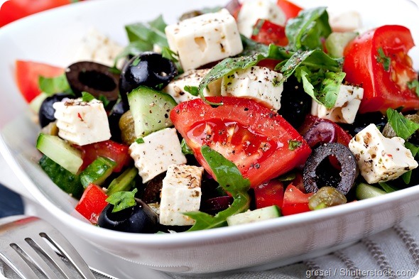
"Whales and other deep-diving marine mammals can pack 10-20 times more myoglobin into their cells than humans can, and that allows them to 'download' oxygen directly into their skeletal muscles and stay active even when they are holding their breath," said Olson, Rice's Ralph and Dorothy Looney Professor of Biochemistry and Cell Biology. "The reason whale meat is so dark is that it's filled with myoglobin that is capable of holding oxygen. But when the myoglobin is newly made, it does not yet contain heme. We found that the stability of heme-free myoglobin is the key factor that allows cells to produce high amounts of myoglobin."
That's important to Olson because he wants to create a strain of bacteria that can generate massive quantities of another protein that's closely related to myoglobin. Olson has spent two decades studying hemoglobin, a larger, more complex oxygen-carrying protein in blood. Olson's goal is to create synthetic blood for use in transfusions. Hospitals and trauma specialists currently rely on donated whole blood, which is often in short supply and has a limited storage life. A crucial part of Olson's plan is maximizing the amount of hemoglobin that a bacterium can express.
"Our results confirm that protein stability is the key," Olson said. "In this study, Premila and George developed an in vitro method for testing myoglobin expression outside of living cells. That allowed us to carefully control all the variables. We found that the amount of fully active myoglobin expressed was directly and strongly dependent on the stability of the protein before it bound the heme group."
All proteins have a characteristic shape, and the globin family of proteins is shaped around a pocket where heme is stored. The heme pocket opens and closes -- much like the pocket of a baseball glove -- to trap and release oxygen.
Samuel, a graduate student in the Department of BioSciences at Rice, said the heme-free form of myoglobin that she studied is called apoprotein or apomyoglobin.
"The more stable the apoprotein, the more final product we could make," she said. "Human apomyoglobin isn't very stable at all compared to that of the diving mammals, which have versions of the apoglobin that are up to 60 times more stable than ours."
Samuel said the stability differences aren't obvious if one simply compares the overall structures of the myoglobin from each species. Their overall shapes, including the shapes of their heme pockets, are the same. However, thanks to subtle differences in their amino acid sequences, the more stable myoglobins are better able to retain their shapes. Samuel said this underlying stability only becomes apparent when one studies the heme-free, or "apo" versions of the protein. She measured stability using chemicals that forced the apoproteins to unfold. By carefully measuring the amount of chemical required, she was able to precisely measure stability.
She said her work was made possible by three earlier studies. In 1999, Emily Scott, a graduate student in Olson's lab, noticed that sperm whale apomyoglobin was much more resistant to chemically induced unfolding than the corresponding human or pig apoproteins. Scott wondered if the resistance to unfolding was a trait of deep-diving whales, so she gathered samples from a variety of mammals and confirmed the idea in 2000.
At the same time, study co-author Smith, another of Olson's graduate students, was examining a catalog of 250 mutant sperm-whale apomyoglobins. He noticed that a certain class of mutations in the heme pocket caused the proteins to become extraordinarily stable even though the mutations damaged their ability to bind heme and oxygen.
Finally, in 2013, Michael Berenbrink of Liverpool University and Kevin Campbell of the University of Manitoba noted that deep-diving mammals expressed large amounts of myoglobin in their muscle tissue. Berenbrink and Campbell systematically analyzed the genes and available information for all mammalian myoglobins, including those from deep-diving species, and found that the myoglobins from aquatic mammals had large positive surface charges compared with those from land animals. They hypothesized that the charge differences allowed the aquatic species to pack more myoglobin into their muscle cells.
"I heard Berenbrink present his work, and I wondered whether we should re-investigate Emily's and Lucian's work on expression levels and apoglobin stability," Olson said. "At the time, we were in the process of trying to screen large-scale libraries of hemoglobin mutants to try to select for higher stability and expression as part of our work on evaluating blood substitutes. George had suggested we use a wheat-germ-based cell-free translation system for those screens, and Premila was preparing to test the methods with myoglobin.
"The three of us decided she should conduct her tests on a series of proteins that included myoglobins from humans, pigs and several of the deep-diving mammals that Emily had tested and Berenbrink had examined," Olson said. "We also used Lucian's Ph.D. results to construct three mutants that were far more stable than anything found in nature."
In her tests, Samuel compared the stability and cell-free expression level of myoglobins from humans, pigs, goosebeak whales, gray seals, sperm whales, dwarf sperm whales and the three mutants, which had low heme affinity but were 50 times more stable than apomyoglobins from the whales. The research confirmed that the stability of apoprotein is directly correlated with expression levels. For example, very little pig and human myoglobin could be made in the cell-free system, which yielded 10- to 20-fold higher amounts of whale and mutant myoglobins.
The results of the cell-free study unequivocally verify the expression-stability correlations that had been anecdotally observed in previous work in both mammalian cells and E. coli, Olson said.
"This work is very important for our projects on synthetic blood substitutes and determining the toxicity of acellular hemoglobin," he said. "Premila has laid the groundwork for high-throughput screening of large libraries of hemoglobin variants without the need for purifying milligram quantities of pure protein. This method is a big step forward in our efforts to identify more stable recombinant hemoglobins."