KNOWLEDGE BAG OF BIOCHEMISTRY

Sunday 16 August 2015

RIBOSOMES CAN TRANSLATE 'UNTRANSLATED REGION' OF Messenger RNA. An Alert for Biologists


In what appears to be an unexpected challenge to a long-accepted fact of biology, Researchers say they have found that ribosomes -- the molecular machines in all cells that build proteins -- can sometimes do so even within the so-called untranslated regions of the ribbons of genetic material known as messenger RNA (mRNA).

whether the proteins made in this unusual way have useful or damaging functions and under what conditions, questions that have the potential to further our understanding of cancer cell growth and how cells respond to stress.
In a summary of the findings in yeast cells, to be published Aug. 13 in the journal Cell, Green and her team report that the atypical protein-making happens when ribosomes fail to get "recycled" when they reach the "stop" signal in the mRNA. For reasons not yet understood, Green says, "rogue" ribosomes restart without a "start" signal and make small proteins whose functions are unknown.
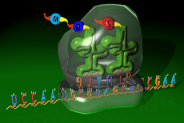
Ribosomes are made out of specialized RNA molecules (DNA's chemical cousin) that work together with proteins to read instruction-bearing mRNAs and "translate" their message to create proteins. Each mRNA begins with a "start" code, followed by the blueprint for a specific protein, followed by a "stop" code. And then there's a segment of code that has always been called the "untranslated region," because scientists never saw it translated into protein.
But no longer, according to Green and postdoctoral fellow Nicholas Guydosh, Ph.D., who, along with a team at the National Institute of Child Health and Human Development, began the project out of curiosity about a yeast protein called Rli1.
Previous studies had shown that Rli1 can split ribosomes into their two component parts once they encounter a stop code and are no longer needed. This "recycling" process, they say, disengages a ribosome from its current mRNA molecule so that it's available to translate another one. But it was unclear whether Rli1 behaved the same way in live cells.
To find out, the researchers deprived living yeast cells of Rli1, predicting that translation would slow down as ribosomes piled up at stop codes. To "see" where the ribosomes were, the team added an enzyme to the cells that would chew up any exposed RNA. The RNA bound by ribosomes would be protected and could then be isolated and identified. As predicted, the depletion of Rli1 increased the number of ribosomes sitting on stop codes. But they also saw evidence of ribosomes sitting in the untranslated region, which they called a surprise.
To find out if the ribosomes were actually reading from the untranslated region to create proteins, the team inserted genetic code in that region for a protein whose quantity they could easily measure. Cells with Rli1 didn't make the protein, but cells missing Rli1 did, proving that their ribosomes were indeed active in the untranslated region.
Further experiments showed that the ribosomes weren't just continuing translation past the stop code to create an extra-long protein. They first released the regularly coded protein as usual and then began translation again nearby.
"It seems like the ribosomes get tired of waiting to be disassembled and decide to get back to work," says Guydosh. "The protein-making work that appears right in front of them is in the untranslated region."
As noted, the purpose of these many small proteins is unknown, but Green says one possibility stems from the fact that ribosomes increase in the untranslated region when yeast are stressed by a lack of food. "It's possible that these small proteins actually help the yeast respond to starvation, but that's just a guess," she says.
Because ribosomes are essential to create new proteins and cell growth, Green notes, scientists believe the rate at which cells replicate is determined, at least in part, by how many ribosomes they have. Cells lacking Rli1 can't grow because their ribosomes are all occupied at stop codes and in untranslated regions. Thus cancer cells increase their levels of Rli1 in order to grow rapidly.
"We didn't understand previously how important ribosome recycling is for the proper translation of mRNA," says Green. "Without it, ribosomes are distracted from their usual work, which is crucial for normal cell maintenance and growth. This finding opens up questions we didn't even know to ask before."

Tuesday 11 August 2015

INTELLIGENT BACTERIA USED FOR DETECTING DISEASES...

Research teams have transformed bacteria into "secret agents" that can give warning of a disease based solely on the presence of characteristic molecules in the urine or blood. To perform this feat, the researchers inserted the equivalent of a computer programme into the DNA of the bacterial cells. The bacteria thus programmed detect the abnormal presence of glucose in the urine of diabetic patients. This work, published in the journal Science Translational Medicine, is the first step in the use of programmable cells for medical diagnosis.


Bacteria have a bad reputation, and are often considered to be our enemies, causing many diseases such as tuberculosis or cholera. However, they can also be allies, as witnessed by the growing numbers of research studies on our bacterial flora, or microbiota, which plays a key role in the working of the body. Since the advent of biotechnology, researchers have modified bacteria to produce therapeutic drugs or antibiotics. In this novel study, they have actually become a diagnostic tool.
Medical diagnosis is a major challenge for the early detection and subsequent monitoring of diseases. "In vitro" diagnosis is based on the presence in physiological fluids (blood and urine, for example) of molecules characteristic for a particular disease. Because of its noninvasiveness and ease of use, in vitro diagnosis is of great interest. However, in vitro tests are sometimes complex, and require sophisticated technologies that are often available only in hospitals.
This is where biological systems come into play. Living cells are real nano-machines that can detect and process many signals and respond to them. They are therefore obvious candidates for the development of powerful new diagnostic tests. However, they have to be provided with the appropriate "programme" for them to successfully accomplish the required tasks.
To do this, Jérôme Bonnet's team in Montpellier's Centre for Structural Biochemistry (CBS) had the idea of using concepts from synthetic biology derived from electronics to construct genetic systems making it possible to "programme" living cells like a computer.
The transcriptor: the cornerstone of genetic programming
The transistor is the central component of modern electronic systems. It acts both as a switch and as a signal amplifier. In informatics, by combining several transistors, it is possible to construct "logic gates," i.e. systems that respond to different signal combinations according to a predetermined logic. For example, a dual input "AND" logic gate will produce a signal only if two input signals are present. All calculations completed by the electronic instruments we use every day, such as smartphones, rely on the use of transistors and logic gates.
This image shows the principle of the use of modified bacteria for medical diagnosis.
During his postdoctoral fellowship at Stanford University in the United States, Jérôme Bonnet invented a genetic transistor, the transcriptor.
The insertion of one or more transcriptors into bacteria transforms them into microscopic calculators. The electrical signals used in electronics are replaced by molecular signals that control gene expression. It is thus now possible to implant simple genetic "programmes" into living cells in response to different combinations of molecules .
In this new work, the teams led by Jérôme Bonnet (CBS, Inserm U1054, CNRS UMR5048, Montpellier University), Franck Molina (SysDiag, CNRS FRE 3690), in association with Professor Eric Renard (Montpellier Regional University Hospital) and Drew Endy (Stanford University), applied this new technology to the detection of disease signals in clinical samples.
Clinical samples are complex environments, in which it is difficult to detect signals. The authors used the transcriptor's amplification abilities to detect disease markers, even if present in very small amounts. They also succeeded in storing the results of the test in the bacterial DNA for several months.
The cells thus acquire the ability to perform different functions based on the presence of several markers, opening the way to more accurate diagnostic tests that rely on detection of molecular "signatures" using different markers.
"We have standardised our method, and confirmed the robustness of our synthetic bacterial systems in clinical samples. We have also developed a rapid technique for connecting the transcriptor to new detection systems. All this should make it easier to reuse our system," says Alexis Courbet, a postgraduate student and first author of the article.
As a proof of concept, the authors connected the genetic transistor to a bacterial system that responds to glucose, and detected the abnormal presence of glucose in the urine of diabetic patients.
"We have deposited the genetic components used in this work in the public domain to allow their unrestricted reuse by other public or private researchers, " says Jérôme Bonnet.
"Our work is presently focused on the engineering of artificial genetic systems that can be modified on demand to detect different molecular disease markers," he adds. In future, this work might also be applied to engineering the microbial flora in order to treat various diseases, especially intestinal diseases.


HOW A FEMALE X CHROMOSOME IS INACTIVATED??????

Chromosomes differentiate men from women. A woman's somatic cells have two X chromosomes, while a man's carry only one. If both X chromosomes and all of their genes were to be active in women, they would have twice as many copies of the proteins that they produce in men. This would consequently result in a disequilibrium that would disrupt the finely balanced biochemistry of the human body.

Nature ensures this does not happen: one of the X chromosomes is completely and permanently inactivated during a female's early development in the womb. The mechanism responsible for this inactivation is not yet fully understood. However, research into mice has shown that a ribonucleic acid (RNA) molecule called Xist plays a pivotal role in the process. Several hundred copies of this molecule attach themselves to one of the two X chromosomes. Scientists believe that these RNA molecules dock onto other molecules which then inactivate the chromosome. A team of researchers lead by Anton Wutz, Professor of Genetics at ETH Zurich, have now discovered several of these inactivation molecules.
Screening to rescue cells
To this end, scientists used mouse stem cells, which exhibited two particular characteristics. Firstly, like unfertilised egg cells (and in contrast to somatic cells), they had just one instance of each chromosome. Secondly, they were modified to a degree that allowed the scientists to continuously produce the Xist RNA. This led to the inactivation of the single X chromosome and the death of the cells, since the genes needed for their continued survival could no longer be read.
In a large-scale screening experiment using these stem cells, scientists were able to identify which genes were important for X inactivation. It is possible to think of the experiment as a sort of rescue operation for the stem cells that would otherwise have died. Specifically, researchers used a virus to randomly damage individual genes in the genetic material of a large number of stem cells . Virus insertions that destroyed a gene, which was required for Xist RNA to inactivate the X chromosome, the X chromosome was not inactivated, and the corresponding cells survived.
The scientists were thus able to isolate surviving stem cells and identify seven genes that are central to X inactivation. One of them is called Spen. Scientists were already aware that Spen produces a protein which allows it to bind with RNA and essentially prevents the genes from being read. In other experiments, ETH researchers were able to show that if a mouse cell lacks the Spen gene, the proteins responsible for altering chromosome structure are not able to accumulate as efficiently at the X chromosome. ETH Professor Wutz explains that further research is required to understand exactly how this mechanism works and what role the other recently discovered genes play in it.
Research made possible thanks to earlier advances
"Genetic research such as this is extraordinarily complex," says Wutz. For example, a significant body of knowledge about mammalian genetics comes from conclusions yielded by research into drosophilidae (fruit flies), which are a model organism for biology and, in particular, for genetic research. Unlike mammals, however, fruit flies have a different chromosome system that does not include X inactivation. You cannot therefore draw on fruit-fly genetics to find gene candidates in mammals.
According to the professor, methodological advances made in recent years have made his research possible. Research of this type is now possible thanks to stem cells with the simple set of chromosomes, created by Wutz five years ago while he was still at the University of Cambridge.
The ETH researchers published their work in the latest issue of the scientific journal Cell Reports. A British research team also published its findings in the same issue. Using a different method -- RNA interference -- they discovered several of the genes involved in X inactivation. One of them is Spen.
Slight differences in humans
The genes for Xist and Spen are found in humans as well. Thus, as Wutz points out, this research offers us some insight into the human system -- at least at the theoretical level, as mouse genetics cannot be mapped directly to humans.
A few years ago, a team of French researchers postulated that, in addition to Xist, humans also have another system which ensures that the single X chromosome in men and one of the two X chromosomes in women remain active. This activating system does not exist in mice. Due to the interplay of activating and inactivating factors, regulation of X chromosomes in humans might therefore be more complicated than originally thought. Geneticists wanting to understand these processes in detail still have plenty of work ahead of them.

Saturday 8 August 2015

HIV GROWS DESPITE TREATMENT... Experts find


HIV can continue to grow in patients who are thought to be responding well to treatment, according to research.


HIV can continue to grow in patients who are thought to be responding well to treatment, according to research. During treatment for HIV the virus hides in blood cells that are responsible for the patient's immune response. The virus does this by inserting its own genetic information into the DNA of the blood cells, called CD4 Tlymphocytes.


During treatment for HIV the virus hides in blood cells that are responsible for the patient's immune response. The virus does this by inserting its own genetic information into the DNA of the blood cells, called CD4 Tlymphocytes.
The study by the University's Institute of Infection and Global Health measured the levels of integrated HIV in the CD4 cells of patients undergoing uninterrupted treatment for up to 14 years, and compared patients receiving treatment for different lengths of time.
The researchers discovered that the amount of HIV found to be integrated in the CD4 cells was undiminished from year 1 to year 14.
The research demonstrates that whenever a CD4 cell multiplies to produce more cells, it copies itself and also copies the HIV genes. This process -- a sort of silent HIV replication -- means the virus does not need to copy itself, produce new virus particles, and infect new CD4 cells -- but is automatically incorporated at the birth of the cell.
Anti-retroviral therapy is given to HIV patients to stop the production of new virus which prevents the infection and death of CD4 T-lymphocytes and the progression of the disease to full-blown AIDS.
Advances in anti-retroviral therapy over the last 30 years mean that most patients can have their virus suppressed to almost undetectable levels and live a long and healthy life. It had been thought that after many years of successful treatment, the body would naturally purge itself of the virus.
Professor Anna Maria Geretti, who led the study, said: "This research shows that sadly, the HIV virus has found yet another way to escape our treatments.
"We always knew HIV is difficult to suppress completely and that it hides inside CD4 cells, but we always hoped that as the body gradually renews its CD4 cells that the hidden HIV would die out. We were surprised to find that the levels of HIV integrated in the CD4 cells didn't reduce over the 14-year period.
"The good news is that we did not see any worsening over time, but the bad news is that these findings really cast doubt over whether HIV can be 'cured' by increasing immune cell responses against it -- a strategy that now looks like it will eventually fail.

Sunday 2 August 2015

WHY WE CAN'T LIVE ON VENUS AND LIVE ON EARTH???

Compared to its celestial neighbours Venus and Mars, Earth is a pretty habitable place. So how did we get so lucky? A new study sheds light on the improbable evolutionary path that enabled Earth to sustain life.

Researchers with the University of British Columbia and University of California, Santa Barbara say that the early loss of these two elements ultimately determined the evolution of Earth's plate tectonics, magnetic field and climate.
"The events that define the early formation and bulk composition of Earth govern, in part, the subsequent tectonic, magnetic and climatic histories of our planet, all of which have to work together to create the Earth in which we live," said Mark Jellinek, a professor in the Department of Earth, Ocean & Atmospheric Sciences at UBC. "It's these events that potentially differentiate Earth from other planets."
On Earth, shifting tectonic plates cause regular overturning of Earth's surface, which steadily cools the underlying mantle, maintains the planet's strong magnetic field and stimulates volcanic activity. Erupting volcanoes release greenhouse gases from deep inside the planet and regular eruptions help to maintain the habitable climate that distinguishes Earth from all other rocky planets.
Venus is the most similar planet to Earth in terms of size, mass, density, gravity and composition. While Earth has had a stable and habitable climate over geological time, Venus is in a climate catastrophe with a thick carbon dioxide atmosphere and surface temperatures reaching about 470 C. In this study, Jellinek and Matt Jackson, an associate professor at the University of California, explain why the two planets could have evolved so differently.
"Earth could have easily ended up like present day Venus," said Jellinek. "A key difference that can tip the balance, however, may be differing extents of impact erosion."
With less impact erosion, Venus would cool episodically with catastrophic swings in the intensity of volcanic activity driving dramatic and billion-year-long swings in climate.
"We played out this impact erosion story forward in time and we were able to show that the effect of the conditions governing the initial composition of a planet can have profound consequences for its evolution. It's a very special set of circumstances that make Earth."
Related image

Saturday 1 August 2015

"Cell aging slowed by putting brakes on noisy transcription''

Working with yeast and worms, researchers found that incorrect gene expression is a hallmark of aged cells and that reducing such "noise" extends lifespan in these organisms. The team published theirfindings this month in Genes & Development
Gene expression is regulated by chemical modifications on chromatin -- histone proteins tightly associated with DNA. Certain chemical groups on histones allow DNA to open up, and others to tighten it. These groups alter how compact DNA is in certain regions of the genome, which in turn, affect which genes are available to be made into RNA (a process called transcription) and eventually proteins.
"Researchers have just started to appreciate how these epigenetic histone modifications may be playing essential roles in determining lifespan," said Berger. She has been studying such epigenetic markings for over two decades and was among the first to pinpoint specific histone modifications that not only are altered during aging, but also directly determine longevity.


"In this study, we found that a type of abnormal transcription dramatically increases in aged cells and that its reduction can prolong lifespan," said Dang, who initiated this line of research while working in Berger's laboratory. "This longevity effect is mediated through an evolutionarily conserved chemical modification on histones. This is the first demonstration that such a mechanism exists to regulate aging."
"We used budding yeast, a single-cell organism, to study the epigenetic regulation of aging and this simple model turned out to be quite powerful," explained Sen. In yeast, aging is measured by the number of times a mother cell divides to form daughters before it stops. This number -- a mean of 25 divisions -- is under tight control and can be either reduced or increased by altering histone modifications, as the researchers found. They showed that when fewer chemical groups of a certain type attach to yeast histones, the abnormal transcription greatly increases in old cells. In contrast, the team found that in yeast strains with a certain enzyme deletion, this abnormal transcription is reduced and lifespan is extended by about 30 percent.
"We have started investigating whether such a longevity pathway can also be demonstrated in mammalian cells," says Berger. "However, these investigations are confounded by the complexity of the genome in more advanced organisms. One of our long-term goals is to design drugs that can help retain these beneficial histone modifications and extend healthy lifespan in humans."