KNOWLEDGE BAG OF BIOCHEMISTRY

Wednesday 10 June 2015

ALTERED GENE LEAVES PEOPLE TOTALLY PAINFREE...

sensing pain

Naturally occurring changes in a previously unstudied gene can prevent people from experiencing pain. And that’s not good. It can leave them dangerously unaware of harm.
Researchers presented the finding May 25 in Nature Genetics.


The gene’s name is PRDM12. Pain is the body’s way of signaling that something is wrong. Certain mutations — naturally occurring changes — in that gene keep people from feeling pain. Robbed of this warning, affected people may fail to protect themselves from unintentional injuries. Such injuries could range from skin burns or scratched eyes to missing digits.
However, there is some good news in the finding. Better understanding of how a mutatedPRDM12 gene blocks pain might one day lead to better treatments for people who suffer from too much pain.
“It’s promising, but there’s a long way to go,” says Simon Halegoua. He’s a neuroscientist at Stony Brook University in New York who did not work on the new study. 
Scientists already knew that mutations in another gene caused a similar birth defect: this insensitivity to pain. In their new study, Geoff Woods of the University of Cambridge in England and his colleagues identified 11 families with mutated forms of the PRDM12 gene. The gene normally instructs cells on how to make a protein that helps pain-sensing nerve cells develop. These pain-warning nerve cells, or neurons, are called nociceptors (NO-see-SEP-terz). People born with some mutations to PRDM12 lack some nociceptor neurons that carry pain signals to the brain. The new study suggests “You need PRDM12 present to grow your pain neurons,” Woods says.

The PRDM12 protein probably helps direct the creation of nociceptor nerves as a baby develops in the womb, Woods says. But that same protein also may have a job to do even after birth. This hints that the gene plays a role in keeping fully formed pain-sensing neurons working well.
The gene’s protein seem to be present only in pain cells and their precursors, says Woods. And that’s “exciting,” he adds. He says it raises the prospect of one day developing a drug that could ultimately tamp down chronic pain without causing problems elsewhere in the body. “The more we can understand about how these nociceptors work,” he explains, “the more likely we are to have treatments for the vast numbers of people with chronic pain.”

Tuesday 9 June 2015

CHILDREN WITH TBI HAVE POORER SLEEP QUALITY, MORE DAYTIME SLEEPINESS

Children with traumatic brain injuries have poorer sleep and more daytime sleepiness in comparison to healthy children, new research confirms. The children with TBI also had impaired emotional, physical and social functioning when compared to healthy children.


A new study suggests that children with traumatic brain injuries (TBI) have poorer sleep and more daytime sleepiness in comparison to healthy children.
Results show that children with TBI were more likely to experience greater daytime sleepiness, sleep disturbances and a poorer overall sleep quality. The children with TBI also had impaired emotional, physical and social functioning when compared to healthy children.
"We were surprised that children with a TBI experienced persistent increases in daytime sleepiness and decreases in sleep quality compared to healthy children," said principal investigator Kimberly Allen, PhD, RN, assistant professor, Center for Narcolepsy, Sleep and Health Research, Department of Women Children and Family Health Science, at the University of Illinois-Chicago.
The research abstract was published recently in an online supplement of the journal Sleep and will be presented June 8, in Seattle, Washington, at SLEEP 2015, the 29th annual meeting of the Associated Professional Sleep Societies LLC.
The study group comprised 15 children with TBI and 15 healthy children, matched on age, race and maternal education level. Parents of children with TBI and parents of health children completed three surveys related to their child's sleep behaviors and sleep quality: Children's Sleep Habits Questionnaire (CSHQ), Child Sleep Wake Scale (CSWS), and the modified Epworth Sleepiness Scale (ESS).

Brain's reaction to certain words could replace passwords

People may not need to remember those complicated e-mail and bank account passwords for much longer. According to a new study, the way your brain responds to certain words could be used to replace passwords.


 A newly published study in academic journalNeurocomputing, researchers from Binghamton University observed the brain signals of 45 volunteers as they read a list of 75 acronyms, such as FBI and DVD. They recorded the brain's reaction to each group of letters, focusing on the part of the brain associated with reading and recognizing words, and found that participants' brains reacted differently to each acronym, enough that a computer system was able to identify each volunteer with 94 percent accuracy. The results suggest that brainwaves could be used by security systems to verify a person's identity.
According to Sarah Laszlo, assistant professor of psychology and linguistics at Binghamton University and co-author of "Brainprint," brain biometrics are appealing because they are cancelable and cannot be stolen by malicious means the way a finger or retina can.
"If someone's fingerprint is stolen, that person can't just grow a new finger to replace the compromised fingerprint -- the fingerprint for that person is compromised forever. Fingerprints are 'non-cancellable.' Brainprints, on the other hand, are potentially cancelable. So, in the unlikely event that attackers were actually able to steal a brainprint from an authorized user, the authorized user could then 'reset' their brainprint," Laszlo said.
Zhanpeng Jin, assistant professor at Binghamton University's departments of Electrical and Computer Engineering, and Biomedical Engineering, doesn't see brainprint as the kind of system that would be mass-produced for low security applications (at least in the near future) but it could have important security applications.
"We tend to see the applications of this system as being more along the lines of high-security physical locations, like the Pentagon or Air Force Labs, where there aren't that many users that are authorized to enter, and those users don't need to constantly be authorizing the way that a consumer might need to authorize into their phone or computer,"

Some host cell environments make malaria parasites resistant to drugs

A new study shows that the different metabolic states of human host cells provide different growth conditions for Plasmodium parasites. It warns that, as a consequence, drugs that work against one Plasmodium species might fail to be effective against the other.


These are mosquito mid-guts infected with P. berghei oocysts. Left panel shows a mosquito mid-gut infected with wild-type parasites. Right panel shows a mosquito mid-gut infected with mutant parasites lacking malate dehydrogenase, which make fewer oocysts but still are able to transmit the parasite.
Of the two species of Plasmodiumparasites commonly infecting humans, P. vivax grows exclusively in immature red blood cells called reticulocytes. P. falciparum can infect reticulocytes, but it grows primarily in mature red blood cells (called erythrocytes) which make up 99% of red cells in circulation. A study published on June 4th in PLOS Pathogens shows that the different metabolic states of these human host cells provide different growth conditions for the respective parasites--and warn that, as a consequence, drugs that work against one Plasmodium species might fail to be effective against the other.
After their birth in the bone marrow, red blood cells undergo a number of changes to develop into highly specialized oxygen transporters. They expel their nucleus (with its DNA content) before they are released into the blood as reticulocytes. As they mature they get rid of many of their other organelles as well, until they are disk-shaped cells full of hemoglobin, a red protein (which gives blood its color) designed to carry oxygen.
To address whether the two classes of host red blood cells offer different resources for parasite survival, and whether these resources could influence antimalarial drug efficacy, Andy Waters, from the Wellcome Trust Centre for Molecular Parasitology at the University of Glasgow, UK, and colleagues from Glasgow and Melbourne, Australia, undertook a comprehensive biochemical analysis of the metabolites present in reticulocytes on one hand and in mature erythrocytes on the other.
They found that reticulocytes contain elevated levels of many metabolites that could potentially be scavenged by the invading and growing malaria parasite. They also saw a marked overlap in metabolic pathways observed in the reticulocyte and those predicted in the parasite. Such common pathways might be uniquely dispensable to Plasmodium during its growth in reticulocytes while they are essential--and hence a good drug target--for growth in erythrocytes.
To test this hypothesis, the researchers used genetic tools to disrupt some of the overlapping pathways in P. berghei, a species that causes malaria in mice and, similar to P. vivax, has a strong preference for growth in host reticulocytes. They found that indeed such mutant P. berghei strains could grow in mouse reticulocytes (utilizing the host's metabolic products).
Moreover, when the researchers compared the sensitivity to a drug known to target one of the overlapping pathways in a test-tube experiment, they found that P. berghei was considerably less sensitive to the drug than P. falciparum, presumably because the former was able to scavenge the metabolites from their reticulocyte host environment whereas no such external sources were available in the erythrocyte host cells invaded byP. falciparum.
Their data, the researchers say, show that reticulocytes provide a highly enriched host cell environment for Plasmodiumparasites. They suggest that "the availability of the reticulocyte metabolome might reduce or block the efficacy of antimalarial drugs that target parasite metabolism. Furthermore reticulocyte resident P. falciparum may enjoy similar protection, giving rise to the possibility that infections could re-emerge."

Monday 8 June 2015

PUPIL DILATION AND ITS MEANING

Scientists are using pupil measurements to study a wide range of psychological processes and to get a glimpse into the mind.

What do an orgasm, a multiplication problem and a photo of a dead body have in common? Each induces a slight, irrepressible expansion of the pupils in our eyes, giving careful observers a subtle but meaningful signal that thoughts and feelings are afoot.
For more than a century, scientists have known that our pupils respond to more than changes in light. They also betray mental and emotional commotion within. In fact, pupil dilation correlates with arousal so consistently that researchers use pupil size, or pupillometry, to investigate a wide range of psychological phenomena. And they do this without knowing exactly why our eyes behave this way. “Nobody really knows for sure what these changes do,” said Stuart Steinhauer, who directs the Biometrics Research Lab at the University of Pittsburgh School of Medicine.
While the visual cortex in the back of the brain assembles the images we see, a different, older part of our nervous system manages the continuous tuning of our pupil size, alongside other functions—like heart rate and perspiration—that operate mostly outside our conscious control. This autonomic nervous system dictates the movement of the iris, like the lens of a camera, to regulate the amount of light that enters the pupil.
The iris is made of two types of muscle: in a brightly lit environment, a ring of sphincter muscles that encircle and constrict the pupil down to as little as a couple of millimeters across; in the dark, a set of dilator muscles laid out like bicycle spokes, which can expand the pupil up to 8 millimeters—approximately the diameter of a chickpea.
Cognitive and emotional events can also dictate pupil constriction and expansion, though such events occur on a smaller scale than the light reflex, causing changes generally less than half a millimeter. But that’s enough. By recording subjects’ eyes with infrared cameras and controlling for other factors that might affect pupil size, like brightness, color, and distance, scientists can use pupil movements as a proxy for other processes, like mental strain.
Princeton psychologist Daniel Kahneman showed several decades ago that pupil size increases in proportion to the difficulty of the task at hand. Calculate 9 times 13, and you pupils will dilate slightly. Try 29 times 13, and they will widen further and remain dilated until you reach the answer or stop trying. As Kahneman says in his recent book, Thinking Fast and Slow, he could divine when someone gave up on a multiplication problem simply by watching for pupil contraction during the experiment.
“The pupils reflect the extent of mental effort in an incredibly precise way,” Kahneman told the German news magazine Der Spiegel, adding, “I have never done any work in which the measurement is so precise.” When he instructed subjects to remember and recite a series of seven digits, their pupils grew steadily as the numbers were presented one-by-one and shrunk steadily as they unloaded the digits from memory.
Subsequent research found that the pupils of intelligent people (as defined by their SAT scores) dilated less in response to cognitive tasks compared to those of less intelligent participants, possibly indicating a more efficient use of brainpower. 
Scientists have since used pupillometry to assess everything from sleepiness to introversion, race bias,schizophrenia, sexual interest, moral judgment, autism, and depression. And while they haven’t been reading people’s thoughts per se, they’ve come pretty close.
“Pupil dilation can betray an individual’s decision before it is openly revealed,” concluded a 2010 study led by Wolfgang Einhäuser-Treyer, a neurophysicist at The Philipp University of Marburg in Germany. In the study, participants were told to press a button at any point during a 10 second interval, and their pupil size correlated with the timing of their decision. The dilation began about 1 second before they pressed the button and peaked 1 to 2 seconds after.
But are pupils informative outside the lab? Men’s Health Magazine says you can tell when it’s “time to make your move” by watching your date’s pupils, but some skepticism is warranted. “It is unclear to me to what extent this can be exploited in completely unrestrained settings,” Einhäuser-Treyer wrote in an email, pointing out that light conditions could easily interfere with attempts at interpersonal pupillometry.
Other efforts to exploit pupil dilations for purposes beyond scientific research have failed. During the Cold War, Canadian officials tried to develop a device they called the “fruit machine” to detect homosexuality among government employees by measuring how their pupils responded to racy images of women and men. The machine, which never worked, was to aid the government’s purge of gay men and lesbians from the civil service and thereby purportedly reduce their vulnerability to Soviet blackmail.
A pupil test for sexual orientation remains as unlikely as it was in the 1960s. Researchers at Cornell University recently showed that sexual orientation correlated with pupil dilation to erotic videos of their preferred gender, but the trend was only apparent when averaged across subjects, and only for male subjects. While pupillometry shows promise as a noninvasive measure of sexual response, they concluded, “not every participant’s sexual orientation was correctly classified” and “an observable amount of variability in pupil dilation was unrelated to the participant’s sexual orientation.”
Pupillometry also became popular in the advertising industry during the 1970s as a way to test consumers’ responses to television commercials, said Jagdish Sheth, a marketing professor at Emory University. But the practice was eventually abandoned. “There was no scientific way to establish whether it measured interest or anxiety,” Sheth said.
Indeed, pupillometry is limited in its ability to distinguish between the many types of cognitive and emotional processes that can affect pupil dilation. “All we can do is watch the change at the end,” Steinhauer said. “We can't monitor everything going into it.”
Still, he added, our eyes are easy to observe and provide a sensitive indicator of cognitive, emotional, and sensory response, making pupillometry a valuable tool for psychological research. “It's like having an electrode permanently implanted in the brain.”

Sunday 7 June 2015

REPROGRAMMING OF DNA OBSERVED IN HUMAN GERM CELLS FOR FIRST TIME........


A team of researchers has described for the first time in humans how the epigenome -- the suite of molecules attached to our DNA that switch our genes on and off -- is comprehensively erased in early primordial germ cells prior to the generation of egg and sperm. However, the study shows some regions of our DNA -- including those associated with conditions such as obesity and schizophrenia -- resist complete reprogramming.


A team of researchers led by the University of Cambridge has described for the first time in humans how the epigenome -- the suite of molecules attached to our DNA that switch our genes on and off -- is comprehensively erased in early primordial germ cells prior to the generation of egg and sperm. However, the study, published in the journal Cell, shows some regions of our DNA -- including those associated with conditions such as obesity and schizophrenia -- resist complete reprogramming.
Although our genetic information -- the 'code of life' -- is written in our DNA, our genes are turned on and off by epigenetic 'switches'. For example, small methyl molecules attach to our DNA in a process known as methylation and contribute to the regulation of gene activity, which is important for normal development. Methylation may also occur spontaneously or through our interaction with the environment -- for example, periods of famine can lead to methylation of certain genes -- and some methylation patterns can be potentially damaging to our health. Almost all of this epigenetic information is, however, erased in germ cells prior to transmission to the next generation
Professor Azim Surani from the Wellcome Trust/Cancer Research UK Gurdon Institute at the University of Cambridge, explains: "Epigenetic information is important for regulating our genes, but any abnormal methylation, if passed down from generation to generation, may accumulate and be detrimental to offspring. For this reason, the information needs to be reset in every generation before further information is added to regulate development of a newly fertilised egg. It's like erasing a computer disk before you add new data."
When an egg cell is fertilized by a sperm, it begins to divide into a cluster of cells known as a blastocyst, the early stage of the embryo. Within the blastocyst, some cells are reset to their master state, becoming stem cells, which have the potential to develop into any type of cell within the body. A small number of these cells become primordial germ cells with the potential to become sperm or egg cells.
In a study funded primarily by the Wellcome Trust, Professor Surani and colleagues showed that a process of reprogramming the epigenetic information contained in these primordial germ cells is initiated around two weeks into the embryo's development and continues through to around week nine. During this period, a genetic network acts to inhibit the enzymes that maintain or programme the epigenome until the DNA is almost clear of its methylation patterns.
Crucially, however, the researchers found that this process does not clear the entire epigenome: around 5% of our DNA appears resistant to reprogramming. These 'escapee' regions of the genome contain some genes that are particularly active in neuronal cells, which may serve important functions during development. However, data analysis of human diseases suggests that such genes are associated with conditions such as schizophrenia, metabolic disorders and obesity.
Walfred Tang, a PhD student who is the first author on the study, adds: "Our study has given us a good resource of potential candidates of regions of the genome where epigenetic information is passed down not just to the next generation but potentially to future generations, too. We know that some of these regions are the same in mice, too, which may provide us with the opportunity to study their function in greater detail."
Epigenetic reprogramming also has potential consequences for the so-called 'dark matter' within our genome. As much as half of human DNA is estimated to be comprised of 'retroelements', regions of DNA that have entered our genome from foreign invaders including bacteria and plant DNA. Some of these regions can be beneficial and even drive evolution -- for example, some of the genes important to the development of the human placenta started life as invaders. However, others can have a potentially detrimental effect -- particularly if they jump about within our DNA, potentially interfering with our genes. For this reason, our bodies employ methylation as a defence mechanism to suppress the activity of these retroelements.
"Methlyation is effective at controlling potentially harmful retroelements that might harm us, but if, as we've seen, methylation patterns are erased in our germ cells, we could potentially lose the first line of our defence," says Professor Surani.
In fact, the researchers found that a notable fraction of the retroelements in our genome are 'escapees' and retain their methylation patterns -- particularly those retroelements that have entered our genome in our more recent evolutionary history. This suggests that our body's defence mechanism may be keeping some epigenetic information intact to protect us from potentially detrimental effects.

Rabbit virus improves bone marrow transplants, kills some cancer cells......


For patients with blood cancers such as leukemia and multiple myeloma, a bone marrow transplant can be both curative and perilous. It replenishes marrow lost to disease or chemotherapy but raises the risk that newly transplanted white blood cells will attack the recipient's body. Now researcher have found that a rabbit virus can deliver a one-two punch, killing some kinds of cancer cells while eliminating a common and dangerous complication of bone marrow transplants.

For patients with blood cancers such as leukemia and multiple myeloma, a bone marrow transplant can be both curative and perilous. It replenishes marrow lost to disease or chemotherapy but raises the risk that newly transplanted white blood cells will attack the recipient's body.
Now researchers say the myxoma virus, found in rabbits, can do double duty, quelling the unwanted side effects of a bone marrow transplant and destroying cancer cells.
The virus could be especially helpful to patients who have recurring cancer but cannot find a suitable bone marrow donor, said Christopher R. Cogle, M.D., the study's lead investigator and an associate professor in the UF College of Medicine's division of hematology and oncology. Bone marrow transplants from partially matched donors carry about an 80 percent risk of graft-versus-host disease, and the myxoma treatment would address that, Cogle said.
The myxoma virus also could improve bone marrow transplant options among African-Americans and the elderly. Those patients are less likely to find fully matched bone marrow donors, which raises the risk of graft-versus-host disease, according to Cogle.
"Myxoma is one of the best strategies because it is effective but doesn't affect normal stem cells," he said.
During laboratory testing on human cells, the process worked this way: The myxoma virus is attached to a type of white blood cell known as a T-cell. The virus-laden white blood cells can then be delivered as part of a bone marrow transplant from a donor. That's when the virus gets activated and goes to work. It blocks graft-versus-host disease, a complication of bone marrow transplants that can cause problems including skin rash, shortness of breath, abdominal pain, jaundice and muscle weakness. In severe cases, these complications can be fatal. The white blood cells then deliver the myxoma virus to cancer cells, which are killed off by the virus.
The findings were published in the April 22 edition of the journalBlood. After successfully testing the process with human cells, researchers are now studying its effectiveness in a mouse model.
The dual action of the myxoma virus is particularly encouraging, said Grant McFadden, Ph.D., a professor in the UF College of Medicine department of molecular genetics and microbiology. It's the first time that a virus has been shown to simultaneously prevent graft-versus-host disease and kill cancer cells in the laboratory, McFadden said.
The process is known to work on blood-related disorders such as multiple myeloma and acute myeloid leukemia but could someday have broader application for other kinds of cancer, he said. The myxoma virus originates among rabbits in Australia and parts of Europe and is benign to humans.
The discovery might never have happened if not for a chance meeting at a coffee kiosk on the health campus. Cogle and McFadden introduced themselves to each other, which led to a collaboration that has lasted seven years.
"It's one of the benefits of a health research campus like UF," Cogle said. "His virus killed the cancer cells that I grew in my lab and spared normal blood stem cells."
Another crucial part of the research team's work was done by Nancy Villa, Ph.D., a research scientist in the division of hematology and oncology. Villa's findings were crucial to understanding and explaining how myxoma prevents graft-versus-host disease, Cogle said. McFadden credits Villa for finding a way to explain to other scientists how the virus-laden white blood cells can prevent graft-versus-host disease and still be an effective killer of cancer cells. That knowledge will be crucial as the team presses on with its research, McFadden said.
After the initial success with human cells, McFadden is cautiously optimistic that a clinical trial could begin within a year. Before that, researchers need to develop a clinical-grade virus, do safety testing and raise about $1 million for clinical trials. The UF-owned patent on the myxoma process has been licensed to a Houston-based company, which will seek to raise money for clinical trials, Cogle said.

Saturday 6 June 2015

MAJOR NEW STUDY REVEALS THE SIMILARITIES AND DIFFERENCES BETWEEN MICE AND HUMAN.........

artists impression of mouse and genetic information19 November 2014 — Powerful clues have been discovered about why the human immune system, metabolism, stress response, and other life functions are so different from those of the mouse. A new, comprehensive study of the mouse genome by an international group of researchers including Penn State University scientists reveals striking similarities and differences with the human genome. The study may lead to better use of mouse models in medical research.
The findings are reported by the Mouse ENCODE Consortium online on November 19, 2014 and in print on November 20 in the study's main paper in Nature and in several other recent and future publications. They examine the genetic and biochemical programs involved in regulating mouse and human genomes. Ross Hardison, the director of the Huck Institute for Comparative Genomics and Bioinformatics at Penn State University, is the senior corresponding author or co-senior author for four of the five new papers by the consortium, including the paper in Nature.
"We didn't know before these research results that there are a large number of genes with expression levels systematically different between mouse and human," Hardison said. The results offer insights into how gene regulation impacts systems important to the biology of humans and other mammals. The results also provide new information to determine how best to use the mouse as a model for studying human biology and disease, and may help to explain some of the limitations of using the mouse for specific kinds of studies.
"Now we also know which genes have expression patterns that are shared between mouse and humans," Hardison said. "For biological processes using genes with conserved expression patterns, the mouse is an excellent model for certain aspects of human biology."
The scientists also found that, in general, the systems that are used to control gene activity have many similarities in mice and humans, and that the basic structure of these systems has been conserved in both species throughout evolutionary time. The researchers found that differences appeared for specific genes and regulatory elements. "Gene regulation is an equation with many possible solutions," said John Stamatoyannopoulos at the University of Washington, a co-senior author with Hardison of the main Nature paper.
The Mouse ENCODE (ENCyclopedia Of DNA Elements) project is building a comprehensive catalog of functional elements in the mouse genome, and is comparing them to those in the human genome. Such elements include genes that code for proteins, non-protein-coding genes, and regulatory elements that control which genes are turned on or off, and when they are turned on or off. Bing Ren at the University of California, San Diego, also a co-senior author with Hardison of the Nature study, said "This is the first systematic comparison of the mouse and human at the genomic level."
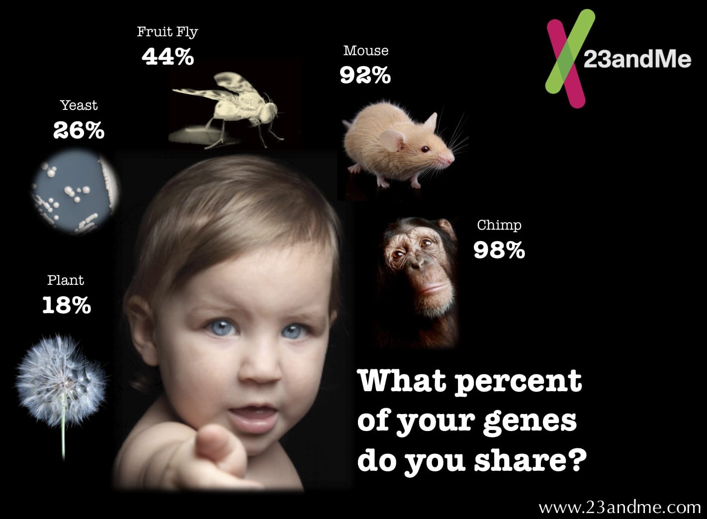
The portion of the Mouse ENCODE effort centered at Penn State focused on comparing mouse and human gene expression and regulatory elements during cell differentiation. This work was done in collaboration with Yu Zhang, associate professor of statistics at Penn State, Feng Yue, assistant professor of biochemistry and molecular biology at Penn State's College of Medicine, and other researchers. "Comparison of the regulatory landscape between mouse and human reveals complex relationships, with some regulatory regions being strictly conserved between mouse and human, other regulatory regions being lost or acquired along each evolutionary lineage -- perhaps reflecting adaptation to different environments, and other regulatory regions being re-used in different tissues," Hardison said. "One would expect that the strictly conserved regulatory regions were particularly important, and this is true, but our collaborative studies have revealed an unexpected basis for their importance." The Mouse ENCODE work revealed that these strictly conserved regulatory regions are active across different tissues, including blood, heart, brain, and others, to a much greater extent than had been previously appreciated. The multiple functions of these regulatory regions may explain the stronger selective pressure during evolution, thus leading to their strict conservation.
The broad, global approaches used in Mouse ENCODE allow investigators to see which genes are expressed in similar patterns and levels between mouse and human, and which ones have divergent patterns. "This information from Mouse ENCODE will enable investigators to make data-driven interpretations of the rich body of information in mouse model systems for potential translation to insights about human biology and health," Hardison said. "For genes with conserved expression patterns, the translation may be quite direct, whereas for others the divergence in expression patterns needs to be incorporated into the inferences for human biology."
More than a dozen companion studies have appeared or will appear in journals including Nature, Nature Communications, Genome Research, and Genome Biology. ENCODE data are freely shared with the biomedical community, and the mouse resource has already been used by researchers outside of ENCODE in about 50 publications.
ENCODE was started with funds from the American Recovery and Reinvestment Act of 2009 and now is supported by the National Human Genome Research Institute (NHGRI), part of National Institutes of Health (NIH). "The mouse has long been a mainstay of biological research models," said NHGRI Director Eric Green, M.D., Ph.D. "These results provide a wealth of information about how the mouse genome works, and a foundation on which scientists can build to further understand both mouse and human biology. The collection of Mouse ENCODE data is a tremendously useful resource for the research community."

It Looks Like An Old Rotten Tree Trunk… But Looking Closer Reveals Something Mind Blowing!

Hey buddy, that’s one gigantic tree trunk… but why is it behind red tape? It looks like it’s all old and rotting with all that funny texture going on. Come closer, you say? Sure… wait… HOLY COW. So apparently this isn’t just a gigantic tree trunk lying around in a gallery. Artist Zheng Chunhui has created a piece called “Along The River During The Quinming Festival” and it seems as though he was on a mission to blow everyone’s mind. Take a look at the photos below and try to imagine the incredible skill and dedication required to create something like this…
Nice tree, buddy… 40-feet long? Pretty impressive! You want me to look closer? Okay…

treetrunk1
Um… WHAT!?
treetrunk2
Okay, so it turns out this is a a wooded carved sculpture. Made with a single tree trunk, it has been recognized as the world’s longest wood carving by the Guinness Book of World Records.
treetrunk4
The sculpture contains over 550 individually carved people. Not to mention all the buildings and foliage.
treetrunk5
It’s is a replica of the famous Chinese painting “Along The River During The Quinming Festival” created by artist Zhang Zeduan during the Song Dynasty.
treetrunk6
This mind-blowing artwork was created by Zheng Chunhui and took four years to complete. Kudos to you, sir.
treetrunk7