

Summary:

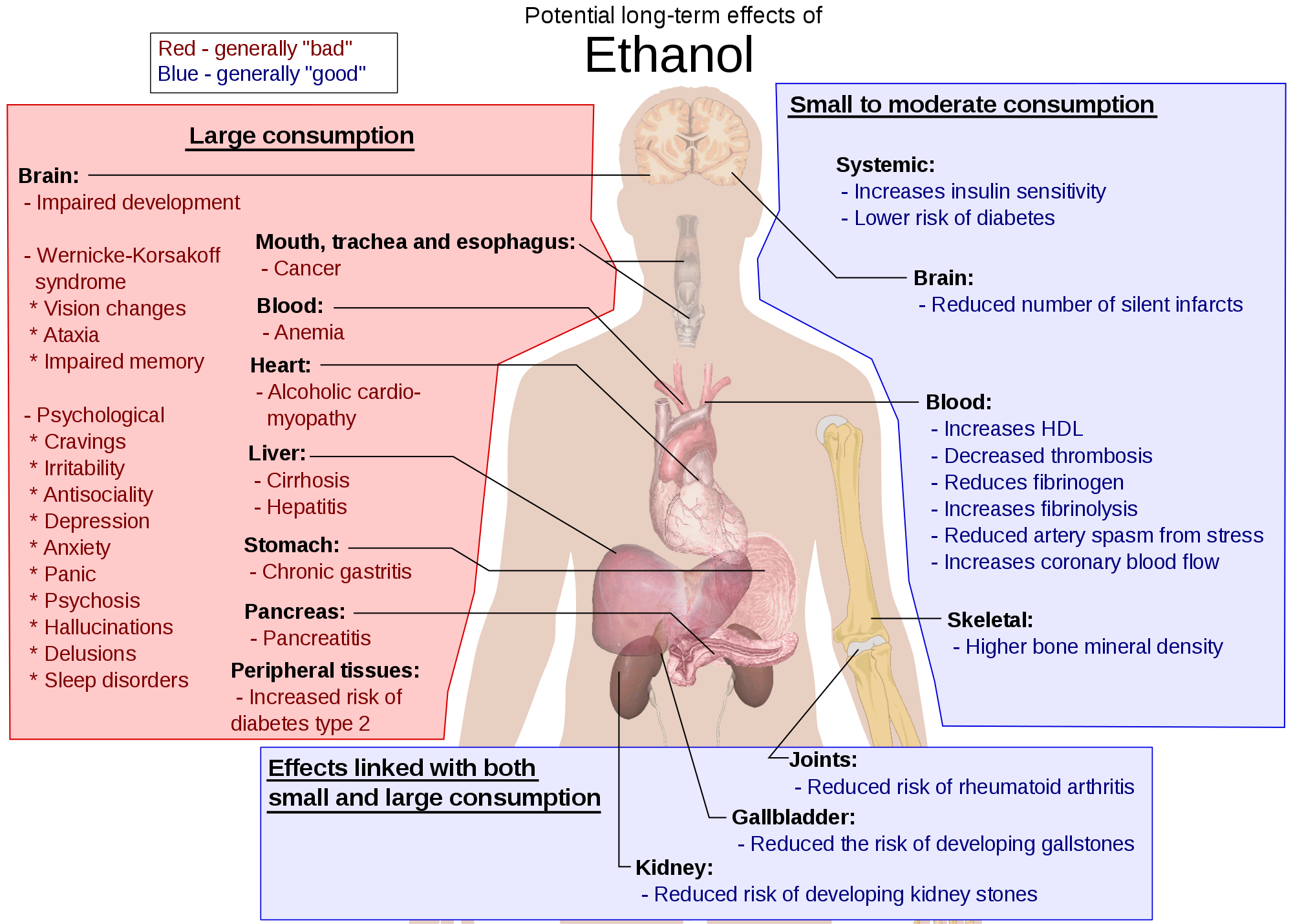 Alcoholism takes a toll on every aspect of a person's life, including skin problems. Now, a new research report helps explain why this happens and what might be done to address it. "The clinical association between alcoholism and severe skin infection is well established," said one expert. "The ability to experimentally model skin immune deficiencies that occur in chronic alcoholics opens up new avenues to test immune-based therapies to better protect this population and thereby limit the spread of infectious disease to the broader community as well."
Alcoholism takes a toll on every aspect of a person's life, including skin problems. Now, a new research report appearing in the April 2015 issue of the Journal of Leukocyte Biology, helps explain why this happens and what might be done to address it. In the report, researchers used mice show how chronic alcohol intake compromises the skin's protective immune response. They also were able to show how certain interventions may improve the skin's immune response. Ultimately, the hope is that this research could aid in the development of immune-based therapies to combat skin infection in people who chronically consume alcohol.
"The clinical association between alcoholism and severe skin infection is well established," said Corey P. Parlet, Ph.D., a researcher involved in the work from the Department of Pathology at the University of Iowa, Carver College of Medicine, Iowa City, Iowa. "The ability to experimentally model skin immune deficiencies that occur in chronic alcoholics opens up new avenues to test immune-based therapies to better protect this population and thereby limit the spread of infectious disease to the broader community as well."
To make their discovery, scientists administered either drinking water consisting of a 20 percent ethanol/water solution or plain water. After 12 weeks on this fluid regimen, with a regular solid food diet, infection outcomes and host defense responses were assessed in mice that were given a skin infection with Staphylococcus aureus (S. aureus). They found that ethanol-consuming mice demonstrated increased illness, including greater weight loss, larger skin lesions and increased bacterial burden. The exacerbation of clinical disease corresponded with an inability to maintain immune cell numbers and activity at the site of infection, especially neutrophils, which are required to heal the infection. Interleukin-17 normally promotes the entry of neutrophils into the skin and their function there. This molecule was reduced in the skin of ethanol-consuming mice. By restoring IL-17 levels, the skin injury in mice was reduced and bacterial clearance defects were improved.
"Co-morbidities associated with chronic alcohol consumption often receive less research attention, yet have significant impact on overall quality of life, healthcare costs and potential infectious disease transmission," said John Wherry, Ph.D., Deputy Editor of the Journal of Leukocyte Biology. "These new studies, together with greater understanding of how to clinically manipulate IL-17 mediated immune responses may lead to new treatment opportunities for alcoholism-associated skin infections.
"
"


.jpg&container=blogger&gadget=a&rewriteMime=image%2F*)


.jpg)
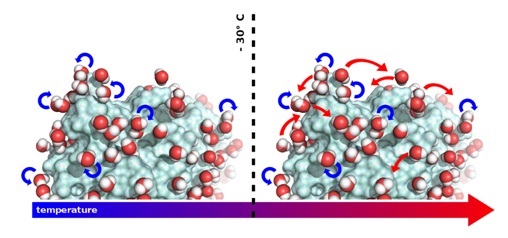 When "visualising" water movement on the protein surface, the team discovered that the molecules rotate around their own axes at temperatures below ‑30°C, temperatures at which proteins are inactive. Above ‑30°C however, whilst continuing to rotate, the water molecules also start to undergo translational diffusion. This is the temperature at which proteins start to be active and the researchers suggest that the capacity of water molecules to "dance" on the surface of proteins enables the dynamics they need to function.


.jpg&container=blogger&gadget=a&rewriteMime=image%2F*)


.jpg)


